Figure 4. Immunohistochemical analysis of Brn-3a. A: Representative slices showing Brn-3a expression in healthy and ischemia/reperfusion (I/R)-injured retinas. Scale bar, 50
μm. B: Brn-3a-positive cells were counted in each section. Following I/R injury, significantly greater numbers of cells were retained
in the retinal ganglion cell (RGC) layer of retinas treated with mutant (triple Y-F) self-complementary adeno-associated virus
type 2 vector encoding brain-derived neurotrophic factor (tm-scAAV2-BDNF) than in those treated with no vehicle (**p<0.01)
or with the tm-scAAV2-GFP vector (**p<0.01; each, n = 3). Bars depict means ± standard deviation (SD).
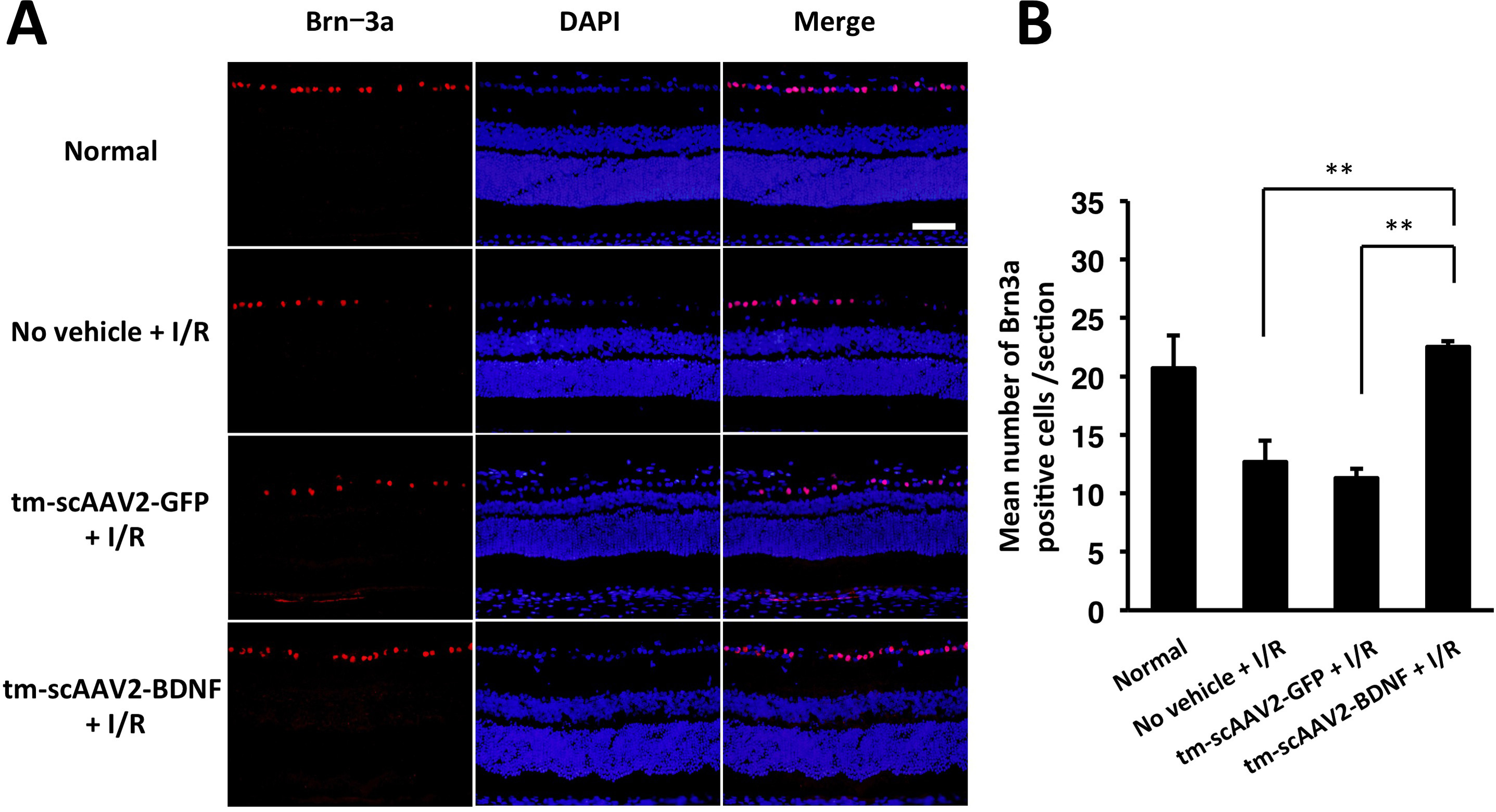
 Figure 4 of
Igarashi, Mol Vis 2016; 22:816-826.
Figure 4 of
Igarashi, Mol Vis 2016; 22:816-826.