Figure 3. Thickness of the inner retina and cell counts in the ganglion cell layer. A: Images of representative slices of healthy retinas and ischemia/reperfusion (I/R)-injured retinas with no vehicle, or with
mutant (triple Y-F) self-complementary adeno-associated virus type 2 vector encoding green fluorescent protein (tm-scAAV2-GFP),
or tm-scAAV2- brain-derived neurotrophic factor (BDNF) treatment. Thickness is defined as the total width between the inner
limiting membrane and the interface of the inner nuclear layer (double arrow). Scale bar, 50 μm. B: Inner retinal thicknesses in each treatment group. n = 6 in each group. Bars depict means ± standard deviation (SD). The
thickness of the I/R-injured retinas treated with the tm-scAAV2-BDNF vector was significantly greater than that of the no
vehicle (**p<0.01) retinas and the retinas treated with the tm-scAAV2-GFP vector (**p<0.01). C: Number of cells in the retinal ganglion cell (RGC) layer or section. A significantly greater number of cells was retained
after I/R injury in the RGC layer of retinas treated with tm-scAAV2-BDNF than in the no vehicle (*p<0.05) retinas or those
treated with the tm-scAAV2-GFP vector (**p<0.01). Bars depict means ± standard deviation (SD).
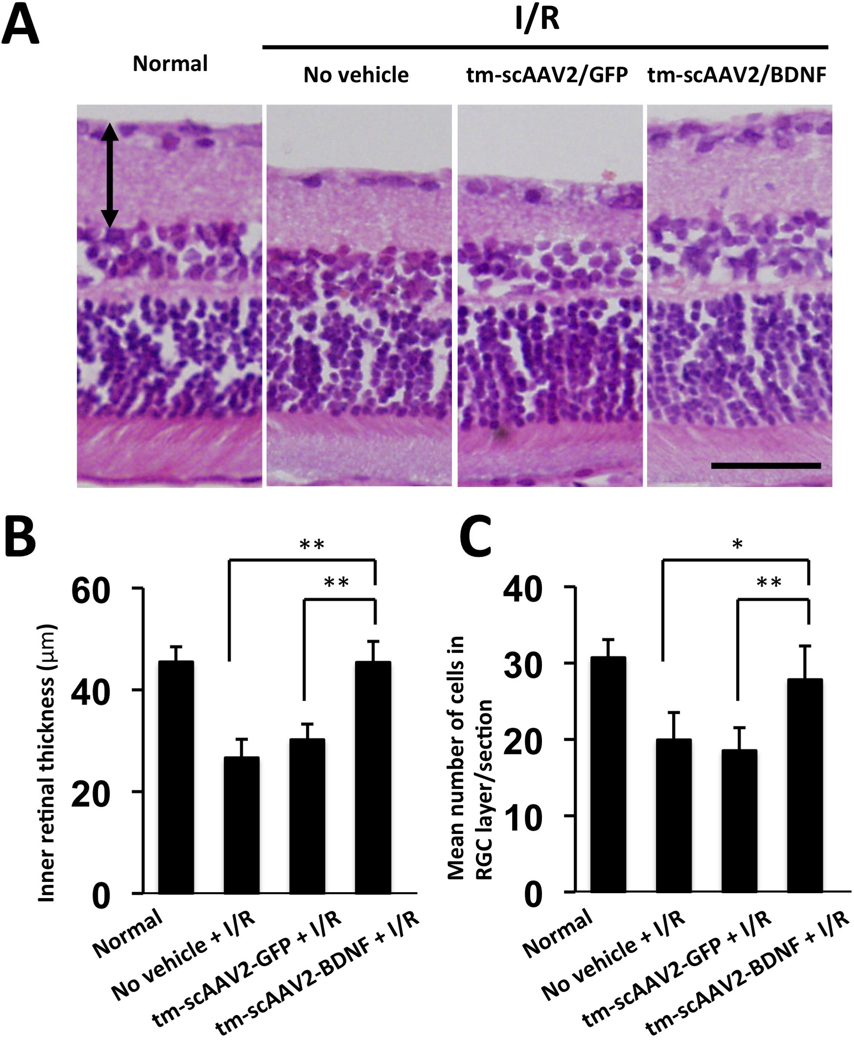
 Figure 3 of
Igarashi, Mol Vis 2016; 22:816-826.
Figure 3 of
Igarashi, Mol Vis 2016; 22:816-826.