Figure 1. Transduction of rat retina with tm-scAAV2-GFP. The upper row shows the green fluorescent protein (GFP) expression pattern
for a healthy retina, and the bottom row shows the GFP expression pattern for the ischemia/reperfusion (I/R) injured retina.
After injection of mutant (triple Y-F) self-complementary adeno-associated virus type 2 vector encoding green fluorescent
protein (tm-scAAV2-GFP) into the rat vitreous cavity, GFP expression was detected in the ganglion cell layer (GCL), Müller,
and inner nuclear layer (INL) cells. Scale bar, 50 μm.
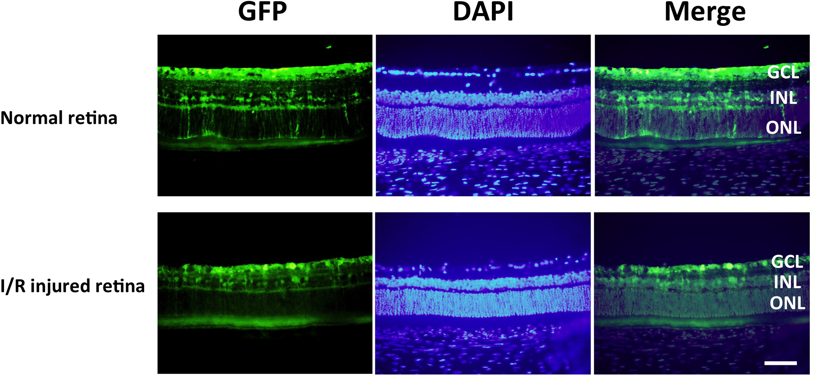
 Figure 1 of
Igarashi, Mol Vis 2016; 22:816-826.
Figure 1 of
Igarashi, Mol Vis 2016; 22:816-826.