Figure 5. Effects of glucose and hypoxia in PEDF secretion by RPE cells. Western blot analysis of pigment epithelium-derived factor
(PEDF) secretion in D407 cells cultured under normoxia (N) and hypoxia (H) conditions and different concentrations of glucose
in the culture medium: 5 mM of D-glucose (corresponding to normoglycemia), 25 mM of D-glucose (corresponding to hyperglycemia),
and mannitol (osmolarity control). n = 4. *p<0.05 represents a significant decrease in PEDF secretion by the RPE cells cultured
under hypoxia with high glucose concentration medium, determined with Tukey’s multiple comparisons test.
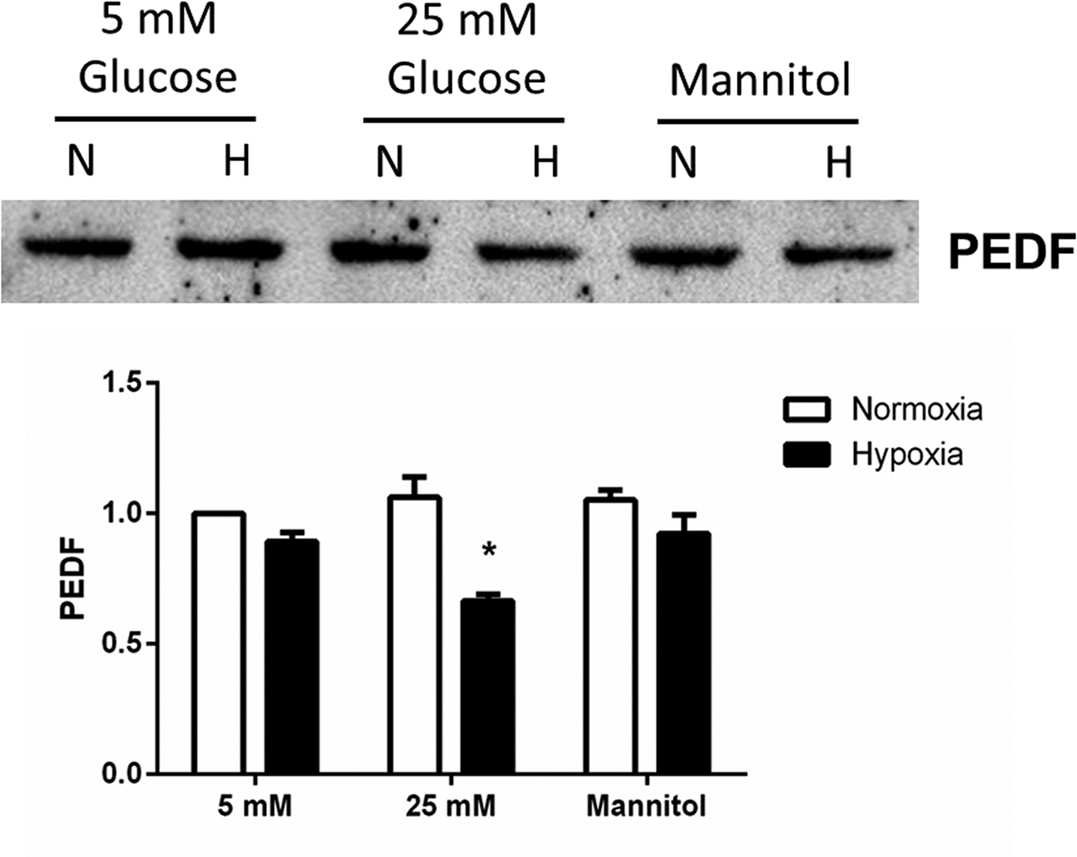
 Figure 5 of
Calado, Mol Vis 2016; 22:761-770.
Figure 5 of
Calado, Mol Vis 2016; 22:761-770.