Figure 1. Findings of retinitis pigmentosa and polydactyly in the male subject. A: Fundus photos demonstrate marked vascular attenuation in both eyes, diffuse RPE atrophic changes with some foveal preservation,
and prominent luteal pigment visualization. B: Loss of the outer retinal layer on optical coherence tomography (OCT) imaging (arrowhead). C: The OCT images show significant retinal thinning in both maculas. D: Post-axial polydactyly of the hands and feet.
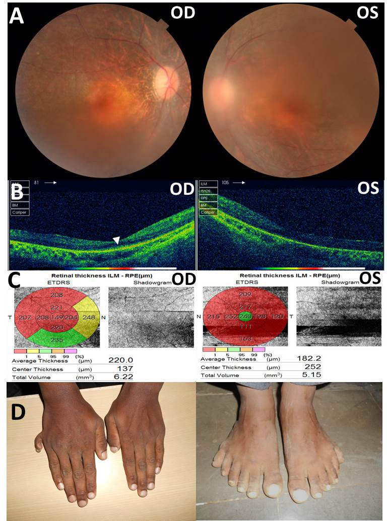
 Figure 1 of
Hulleman, Mol Vis 2016; 22:73-81.
Figure 1 of
Hulleman, Mol Vis 2016; 22:73-81.