Figure 3. EYS variations identified in the present study. A: Schematic representation of the linear location of the identified EYS variants in the context of the genome (upper) and the protein (below). B–E: Crystal structural models of the wild-type (B and D) and mutant protein eyes shut homolog (C and E).
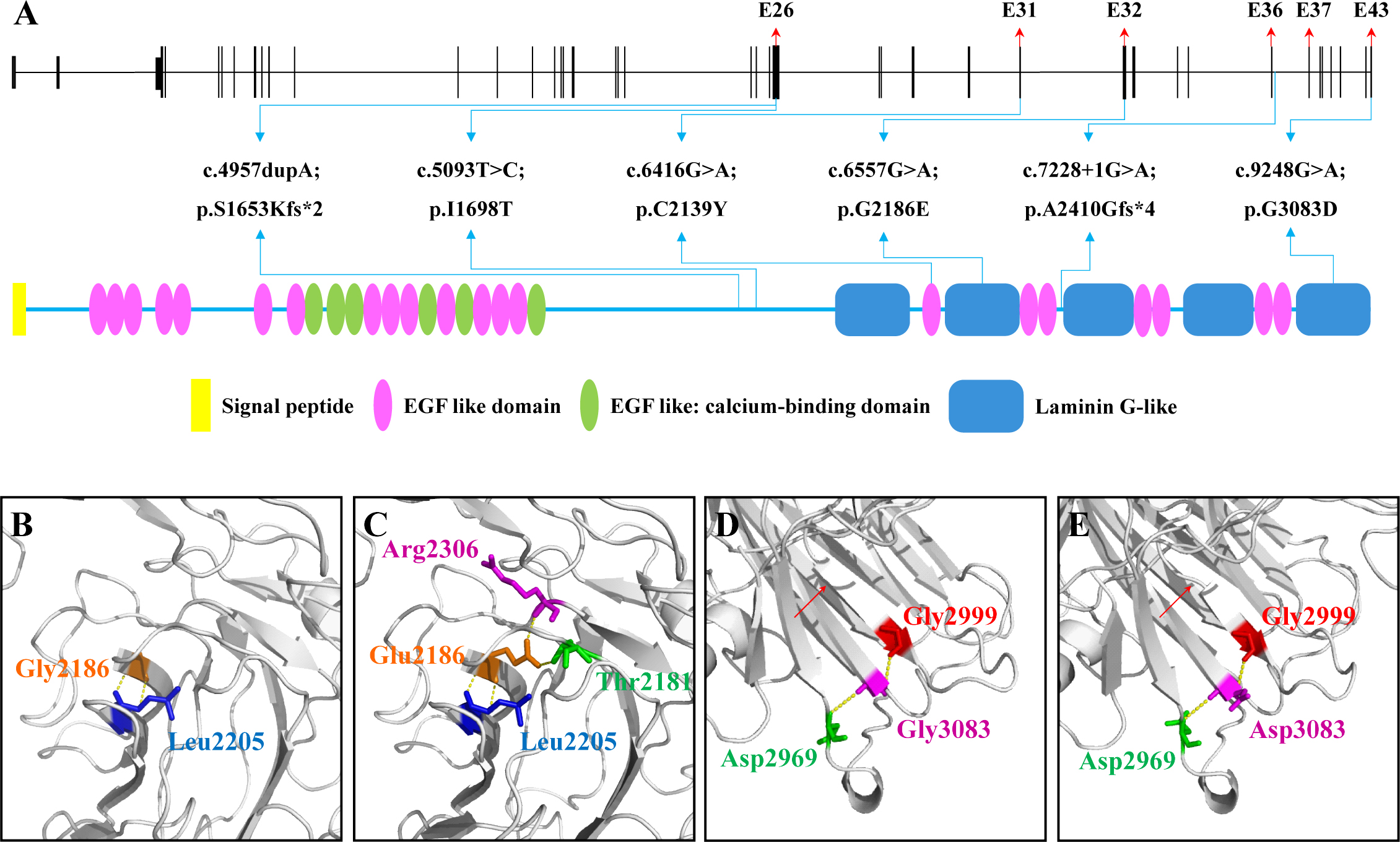
 Figure 3 of
Gu, Mol Vis 2016; 22:646-657.
Figure 3 of
Gu, Mol Vis 2016; 22:646-657.