Figure 2. Fundus and OCT presentations. A and C: Fundus photographs of patient ARRP07-II:1 (A) and ARRP09-II:1 (C) indicate attenuated arterials and waxy optic discs without pigmentation. B and D: Typical retinitis pigmentosa (RP) presentations are revealed in the fundus of patients ARRP08-II:2 (B) and ARRP10-II:3 (D), suggesting arterial attenuation, waxy optic disc, and bone spicule-like pigmentation. E: Fundus of a control individual. F–I: Optical coherence tomography (OCT) presentations of patients ARRP07-II:1 (F), ARRP08-II:2 (G), ARRP09-II:1 (H), and ARRP10-II:3 (I) suggest an attenuated outer nuclear layer (ONL), RPE, and loss of outer/inner segments (IS/OS). The macular regions of all
patients were relatively preserved.
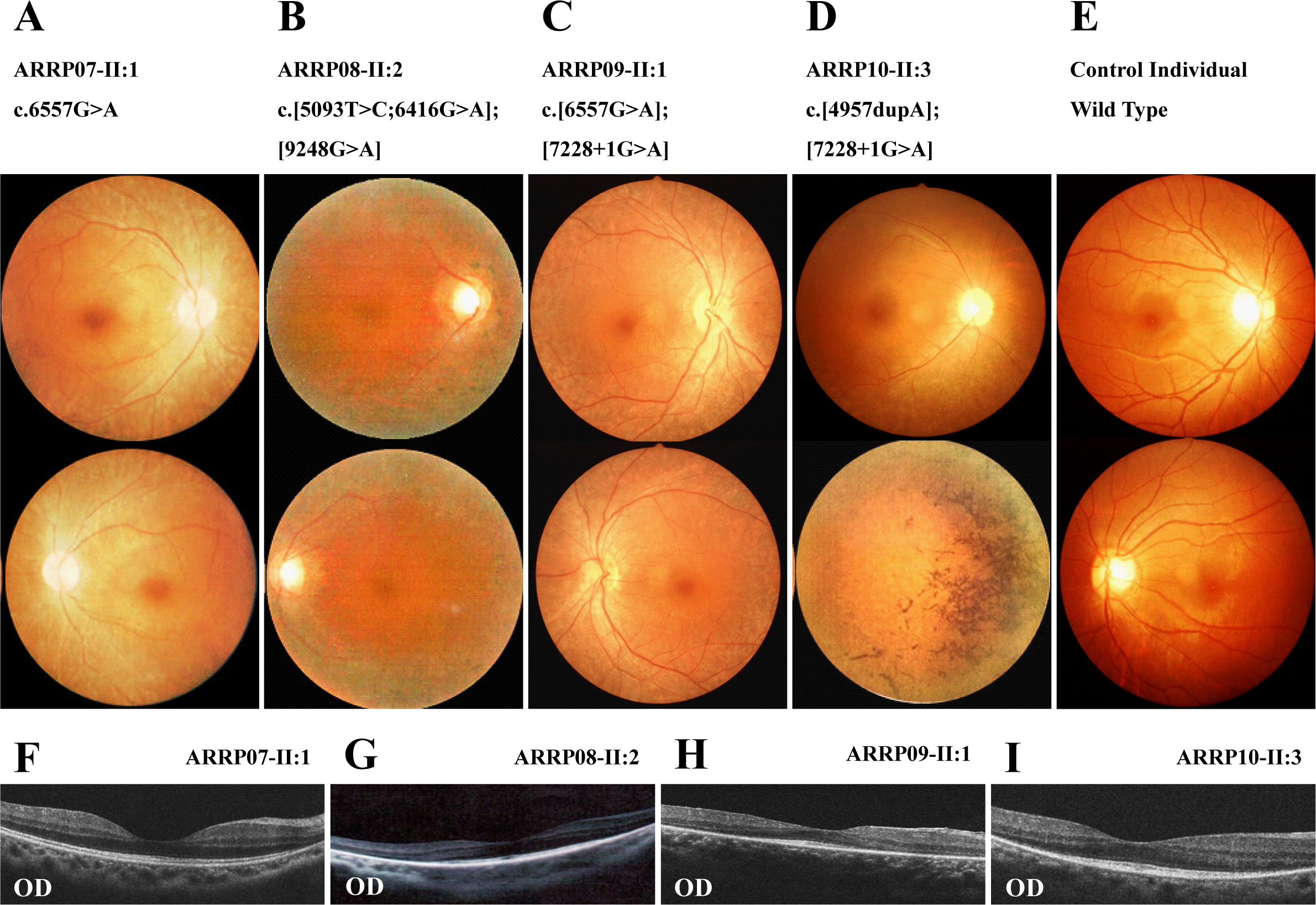
 Figure 2 of
Gu, Mol Vis 2016; 22:646-657.
Figure 2 of
Gu, Mol Vis 2016; 22:646-657.