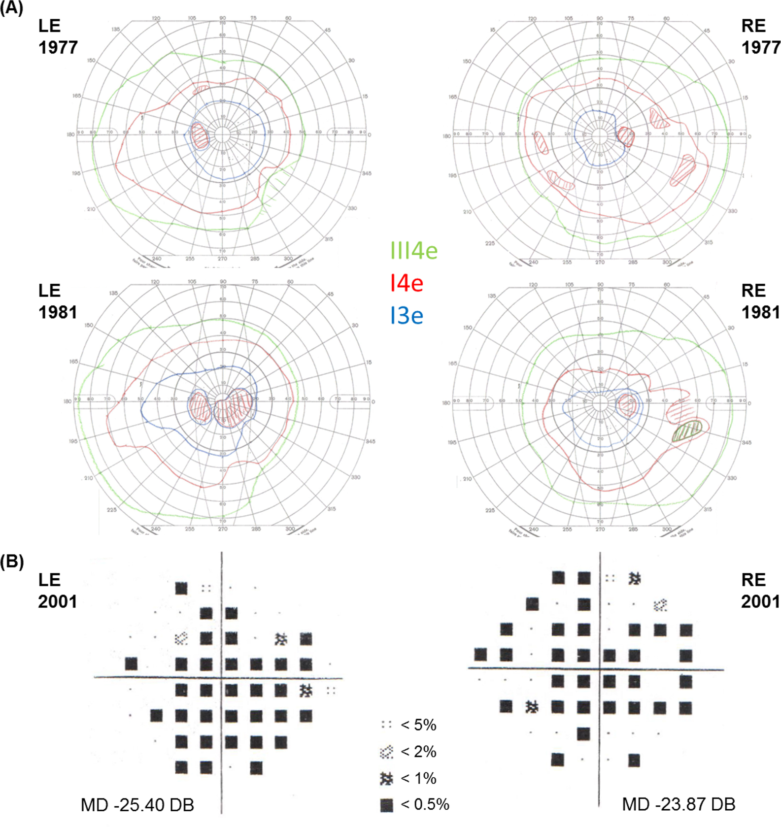
Figure 4. Visual fields for patient 2.2. A: Goldmann visual fields at ages 36 and 40 years of the left eye (LE) and the right eye (RE) demonstrate central scotomas
increasing over time particularly on the LE. Three different light stimulus sizes and intensities, III4e, I4e, and I3e, are
indicated with colored isopters. Light stimulus size III is larger than I, and intensity level 4 is greater than 3. B: Humphrey 24–2 visual fields at age 59 years demonstrate extensive loss of the central field in both eyes. Mean deviation
(MD) indicates the overall average loss of field in decibels (dB). Shaded symbols represent probability indicators of the
statistical likelihood of the field being normal at that location. The darker the symbol, the less likely it is that the field
is normal at that tested location.

 Figure 4 of
Hull, Mol Vis 2016; 22:626-635.
  Figure 4 of
Hull, Mol Vis 2016; 22:626-635.