Figure 2. Fundus photographs of individuals with retinal dystrophy. A: OD and OS of individual 8 (affected, 25 years) of PKRP262. B: OD and OS of individual 12 (affected, 10 years) of PKRP262. C: OD and OS of individual 16 (affected, 12 years) of PKRP358. Fundus photographs of affected individuals show bone spicule-like
pigmentation in the mid-periphery of the retina, attenuated retinal arteriole, severe maculopathy, and disc pallor. OD = oculus
dexter; OS = oculus sinister.
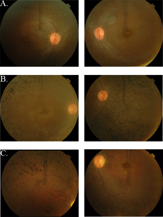
 Figure 2 of
Kabir, Mol Vis 2016; 22:610-625.
Figure 2 of
Kabir, Mol Vis 2016; 22:610-625.