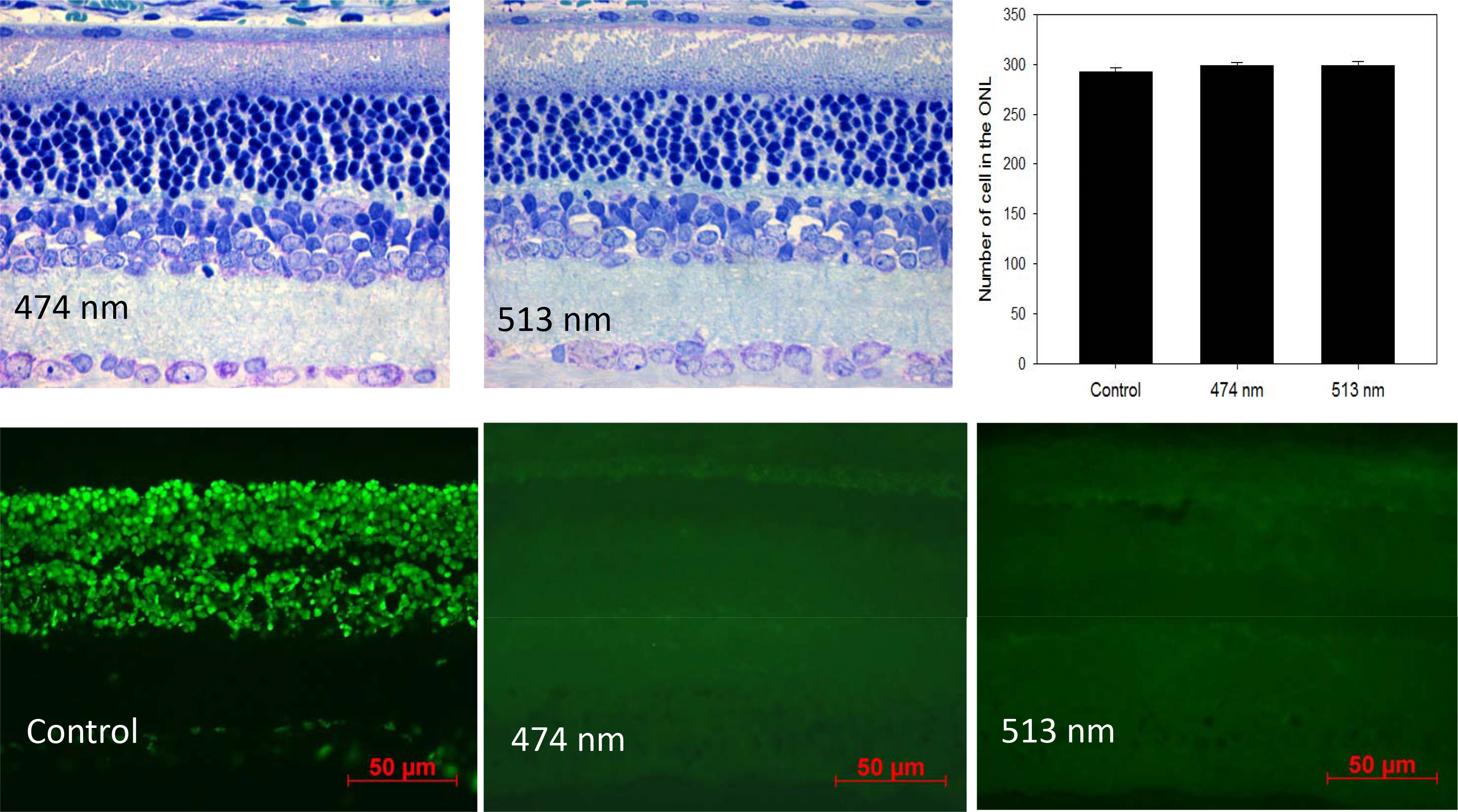
Figure 3. Top panels. The exposure to blue light (λ
max 474), green light (λ
max 513), or fluorescent light at the intensity of 1×10
−1 μW/cm
2 for 4 h/day for 30 days did not produce a significant change in the number of cells in the photoreceptor layers of the Sprague-Dawley
rats (n=6; see [
121] for details about the methods used to quantify cells in the photoreceptor layer). Lower panels. The exposure to blue or
green light-emitting diodes (LEDs) for 4 h in the middle of the day did not induce apoptosis. Terminal deoxynucleotidyl transferase-mediated
uridine 5′-triphosphate-biotin nick end labeling (TUNEL) assay: 4- to 6-week-old Sprague-Dawley rats (n=6) were anesthetized
(75 mg/kg ketamine and 23 mg/kg xylazine), kept on heating pads (37 °C), and exposed to blue or green light for 4 h. The pupils
were dilated with 1% atropine and 2.5% phenylephrine eye drops 45 min before the light exposure. Rats were then killed 16
h after the exposure to blue light or green light. The eyes were explanted and fixed using freshly prepared 4% polyformaldehyde
in PBS, pH 7.4 for 20 min at room temperature. They were washed 3X with PBS, permeabilized with freshly prepared 0.1% Triton
X-100 in 0.1% sodium citrate for 2 min on ice (2–8 °C), and then the TUNEL reaction was performed according to the instructions
included in the manual (In Situ cell Death Detection kit). The slides were incubated in a humidified container for 60 min
at 37 °C in the dark. Slides were rinsed 3X with PBS, and samples were analyzed under a fluorescence microscope (Zeiss Axioskop).
 Figure 3 of
Tosini, Mol Vis 2016; 22:61-72.
Figure 3 of
Tosini, Mol Vis 2016; 22:61-72.