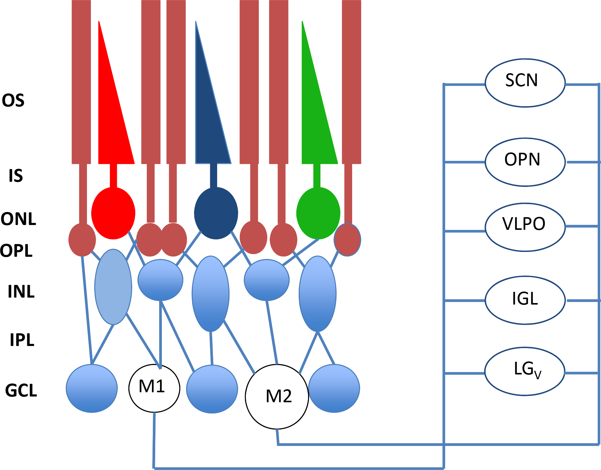
Figure 2. In addition to the classical photoreceptors (rods and cones), ipRGCs are present in the retina. Recent studies have shown
that at least two types of intrinsically photosensitive retinal ganglion cells (ipRGCs) have been identified: M1 and M2. Most
of the M1 cells project to the suprachiasmatic nucleus (SCN) of the hypothalamus whereas the number of M1 and M2 projecting
to the olivary pretectal nucleus (OPN) is similar (55% from M1 cells versus 45% from M2 cells). The M1 cells are considerably
smaller but respond with significantly larger depolarizations and light-induced currents than do the M2 cells. Other neural
targets of ipRGCs not shown in the figure include the preoptic area, sub-paraventricular zone, anterior hypothalamic nucleus,
lateral hypothalamus, medial amygdaloid nucleus, lateral habenula, lateral geniculate nucleus (dorsal division), bed nucleus
of the stria terminalis, periaqueductal gray, and superior colliculus. OS=outer segments; IS=inner segments; ONL=outer nuclear
layer; OPL=outer plexiform layer; INL=inner nuclear layer; IPL=inner plexiform layer; GCL=ganglion cell layer; from [
31] with permission.
 Figure 2 of
Tosini, Mol Vis 2016; 22:61-72.
Figure 2 of
Tosini, Mol Vis 2016; 22:61-72.