Figure 4. Increased number of microglia/macrophages in the retina at 72 h. A: Representative light depicting a significant increase in CX3CR1-GFP-positive microglia and macrophages in the ischemic ipsilateral
eye at 72 h compared to the contralateral side. B: Fluorescence ophthalmoscopic images depicting a significant increase in CX3CR1-GFP-positive microglia and macrophages in
the ischemic ipsilateral eye at 72 h compared to the contralateral side. The green fluorescence signal intensity was measured
in the sham and stroke groups at 72 h after reperfusion (n = 4/group, C). Error bars show mean ± standard error of mean (SEM). Abbreviations: SH = sham, ST = stroke. *p<0.05; **p<0.01; ***p<0.001.
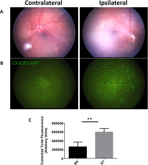
 Figure 4 of
Ritzel, Mol Vis 2016; 22:575-588.
Figure 4 of
Ritzel, Mol Vis 2016; 22:575-588.