Figure 1. Blood flow reduction through the ophthalmic artery via middle cerebral artery occlusion. The middle cerebral artery (MCA)
and the ophthalmic artery in the mouse and occlusion following suture insertion. A: A representative image showing the gross observation of acute sensitivity in the ipsilateral eye during MCA occlusion. B: Ophthalmoscopic imaging depicts blood flow to the contralateral (left) and ipsilateral (right) retina during MCA occlusion.
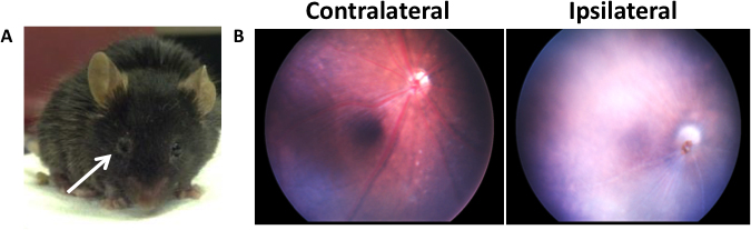
 Figure 1 of
Ritzel, Mol Vis 2016; 22:575-588.
Figure 1 of
Ritzel, Mol Vis 2016; 22:575-588.