Figure 4. Purification and expansion of RPCs. A: Fluorescence-activated cell sorting (FACS) analysis showed Chx10-positive proliferating retinal progenitor cells were further
enriched after manual selection, approximately from 63.70% to 85.87%. Blue line: before selection, green line: after selection.
B: Phase contrast images of P0 retinal progenitor cells (RPCs). Single-cell RPCs have emerged from replated neuroepithelial
clusters and formed a monolayer after 5–6 days. Scale bar = 50 μm. C–D: Confocal images of P0 RPCs stained with early eye field markers Pax6 (C, low magnification; D, high magnification). Scale bar = 50 μm. E: Phase contrast images of established RPC line at passage P6. Neuroepithelial clusters were further dissociated for the single-cell
RPC sub-culture. Scale bar = 50 μm. F–G: Confocal images of dispersed RPCs stained with eye field markers Chx10, Rx, and Ki67. Scale bars = 50 μm. H: Dispersed RPCs were efficiently propagated three- to fourfold once a week on average in the RPC medium. The graph above
shows the mean ± standard deviation (SD; n = 3).
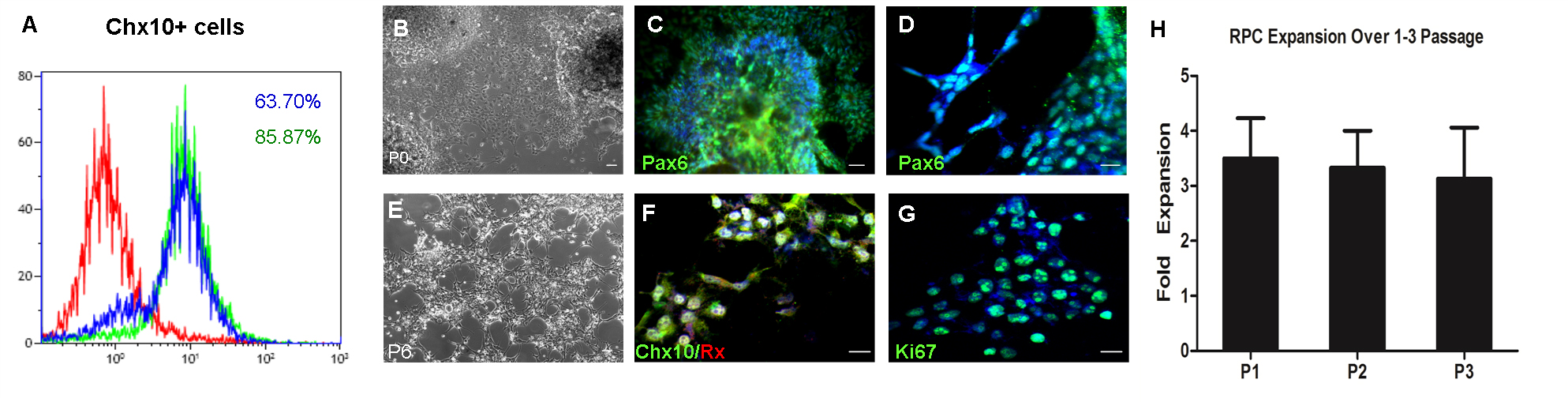
 Figure 4 of
Deng, Mol Vis 2016; 22:536-547.
Figure 4 of
Deng, Mol Vis 2016; 22:536-547.