Figure 6. Immunohistochemistry of mouse and human meibomian gland tissue cryosections. Ten-micron cryosections of optimum cutting temperature
(OCT)-embedded (A) mouse and (B) human eyelid tissue was stained with anti-DKKL1 (red) at a 1/100 dilution and Alexa Fluor 546 secondary antibody at a 1/500
dilution while 4′,6-diamidino-2-phenylindole (DAPI) was used to counter-stain the cell nuclei (green).
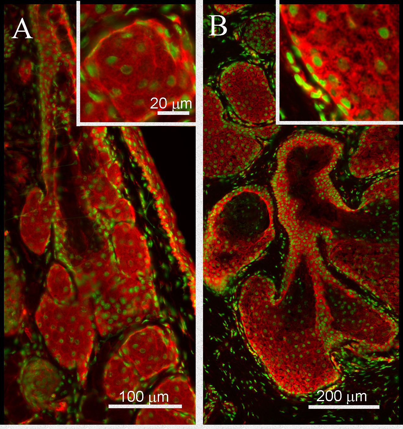
 Figure 6 of
Parfitt, Mol Vis 2016; 22:518-527.
Figure 6 of
Parfitt, Mol Vis 2016; 22:518-527.