Figure 5. Quantitative comparison of collagen orientation between the (A–B) carrier and (C–D) affected ADAMTS10 mutant posterior canine scleras, showing the average percentage of polar vectors circumferentially, radially, or otherwise
oriented (with respect to the optic nerve head) within the peripapillary (PPS) and mid-posterior (MPS) scleral regions.
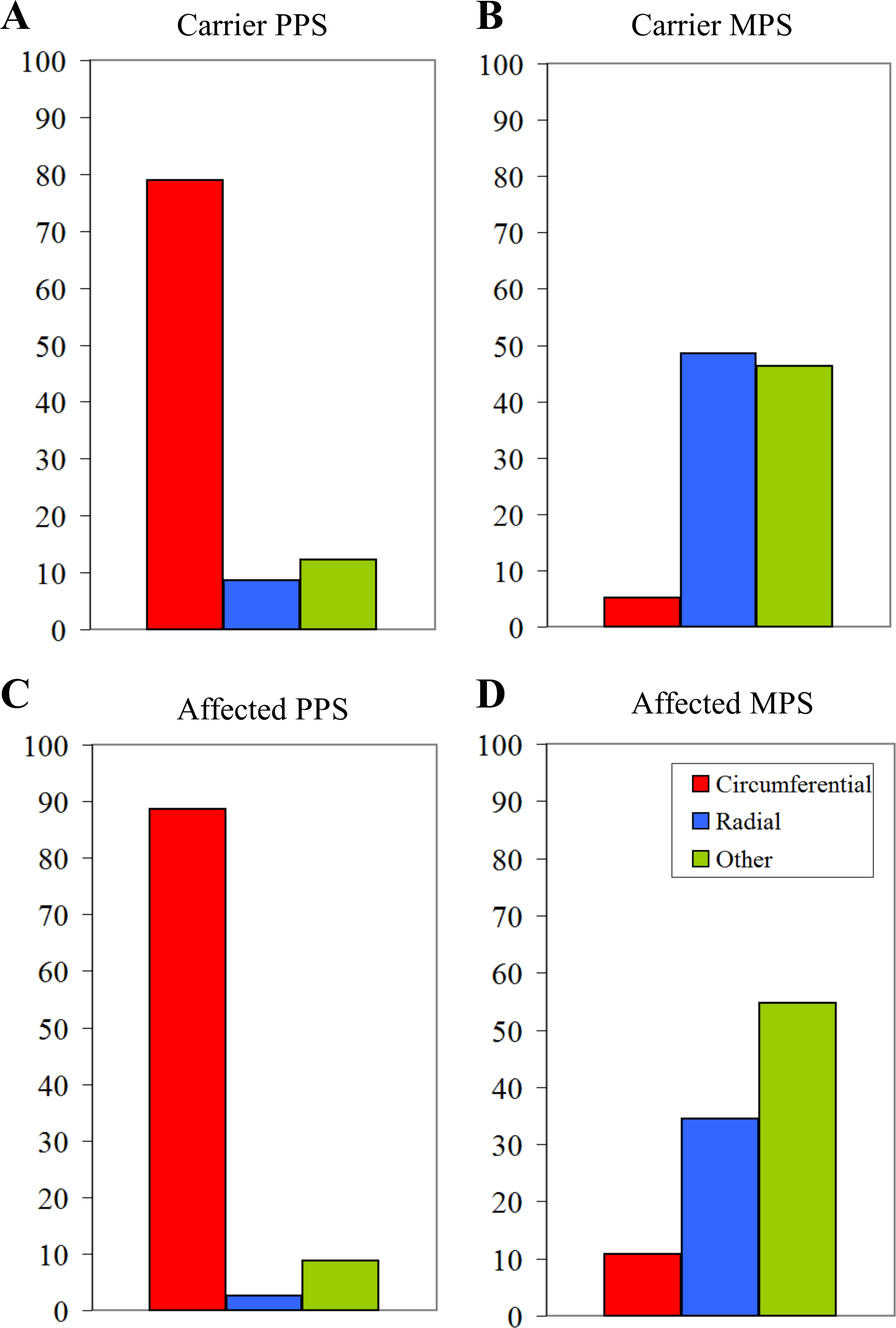
 Figure 5 of
Boote, Mol Vis 2016; 22:503-517.
Figure 5 of
Boote, Mol Vis 2016; 22:503-517.