Figure 4. Collagen orientation maps. Polar vector maps of collagen fibril orientation across the (A–C) carrier and (D–F) affected ADAMTS10 mutant posterior canine scleras. Arrowheads and arrows mark the characteristic features associated with the nasal blood vessel
(long posterior ciliary artery) and peripapillary collagen annulus, respectively. Data sampling interval: 0.4 mm. The plots
have been scaled according to the color key.
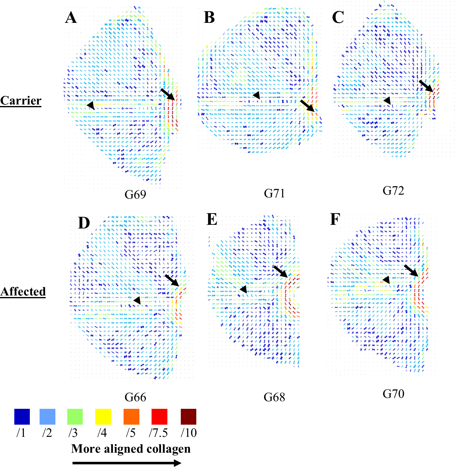
 Figure 4 of
Boote, Mol Vis 2016; 22:503-517.
Figure 4 of
Boote, Mol Vis 2016; 22:503-517.