Figure 3. Production of a collagen fibril orientation map. A: Photograph of posterior canine sclera specimen G69. ONH marks the optic nerve head position. Arrowhead: prominent nasal blood vessel (long posterior ciliary artery) is an anatomic landmark of the canine sclera. B: Montage of the wide-angle X-ray scattering (WAXS) patterns recorded from the specimen at 0.4 mm spatial resolution. C: Expanded view of a WAXS pattern from the peripapillary scleral region. The marked intensity lobing of the collagen intermolecular
peak indicates that collagen is preferentially oriented in the direction of the white arrow. D: Polar vector plot of collagen orientation distribution extracted from the pattern in C. E: Montage of polar vector plots showing collagen orientation across the whole specimen. Arrowhead: An abrupt change from horizontal to near-vertical collagen demarcates the position of the vessel visible in A. Arrow: circumferentially oriented collagen, ringing the optic nerve head (ONH), characterizes the peripapillary sclera. The plots
have been scaled according to the color key.
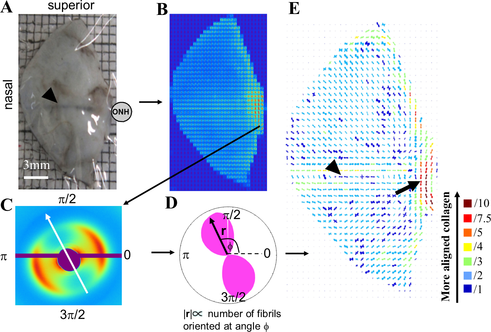
 Figure 3 of
Boote, Mol Vis 2016; 22:503-517.
Figure 3 of
Boote, Mol Vis 2016; 22:503-517.