Figure 4. Fundus photographs and fluorescein angiogram pictures of the patients with FEVR with novel changes (p.H50D, p.G113D, and p.D23EfsX9)
identified in the NDP gene. A: Patient ID: family 33-II:2 (p.H50D); the right eye of the patient shows straightening of the blood vessels and macular dragging
toward the inferotemporal area due to fibrovascular traction. B: Patient ID: family 33-I:2 (p.H50D; affected mother of the proband); the left eye of the patient shows vitreoretinal traction
with macular dragging. C, D: Patient ID: family 72-IV:1 (p.H50D); the right (C) and left (D) eyes show dragging and vitreoretinal traction with an ectopic macula. E: Patient ID: family 139-II:2 (p.H50D); the right eye of the patient shows an avascular peripheral retina with neovascularization
and laser scars after the treatment. F: Patient ID: family 85-II:1 (p.G113D); fundus fluorescein angiogram of the left eye shows an avascular peripheral retina,
straightening of the blood vessels, and dye leakage at the avascular and neovascular junction. G, H: Patient ID: family 21-III:2 (p.D23EfsX9; affected mother of the proband); the right (G) and left (H) eyes of the patient show pigmentation and vitreoretinal traction with a dragged macula.
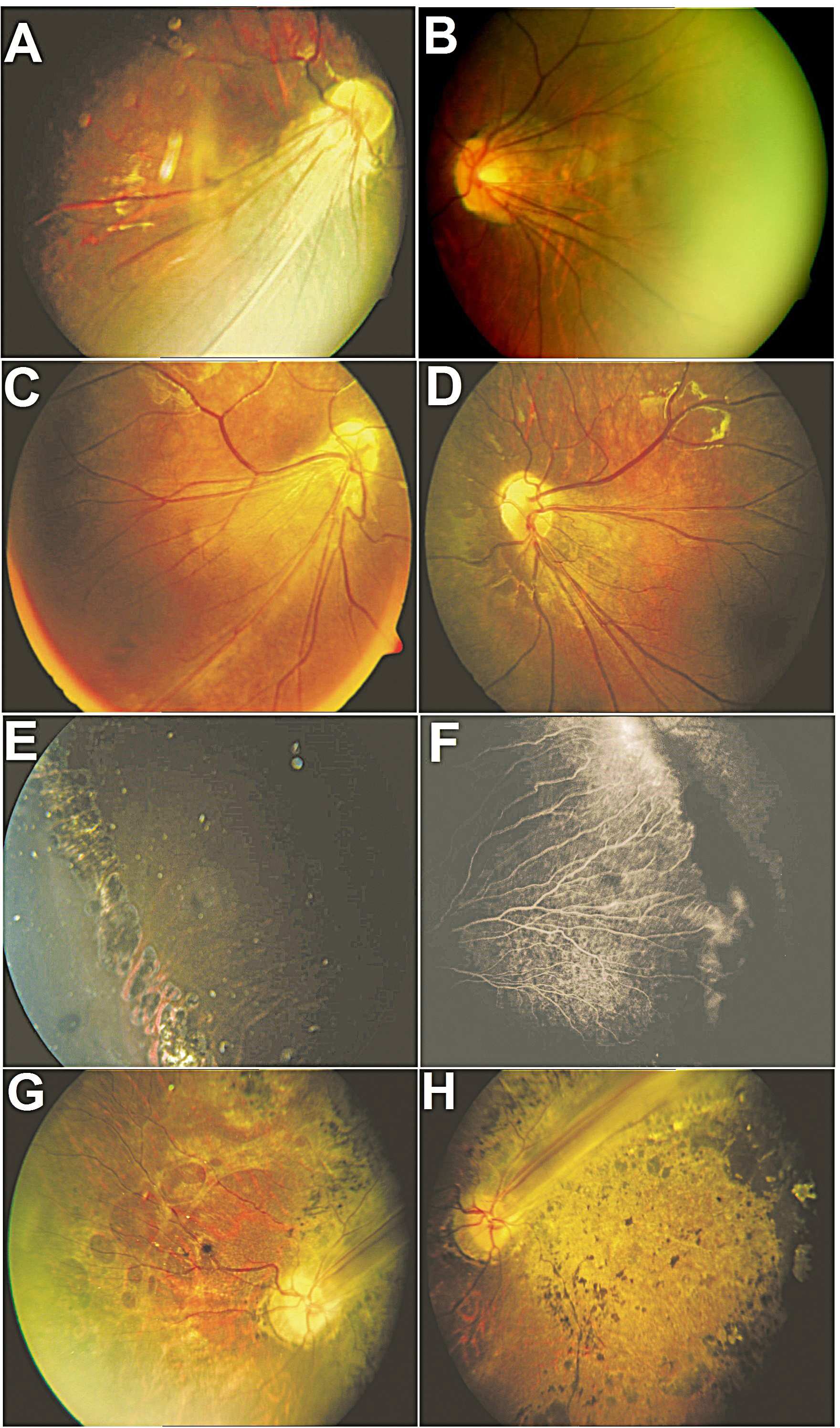
 Figure 4 of
Musada, Mol Vis 2016; 22:491-502.
Figure 4 of
Musada, Mol Vis 2016; 22:491-502.