Figure 3. Western blot showing expression of the mRNA of tenascin-C in vascular smooth muscle cells (VSMCs). A: Expression of the mRNA of tenascin-C normalized to GAPDH in VSMCs stimulated with the indicated concentrations of IL-13.
B: ELISA analysis of tenascin-C in cell lysates and supernatants in VSMCs stimulated with the indicated concentrations of IL-13.
VSMCs stimulated by IL-13 produced and secreted tenascin-C in a dose-dependent manner. C: Western blot analysis of tenascin-C in cell lysates in VSMCs stimulated with the indicated concentrations of IL-13. Mouse
monoclonal anti-tenascin-C clone 4F10TT antibody shows a bond at approximately 300 kDa. β-actin was used as a loading control.
*p <0.001, compared to control. Bars are mean ± standard error of the mean (SEM, n = 4 per group).
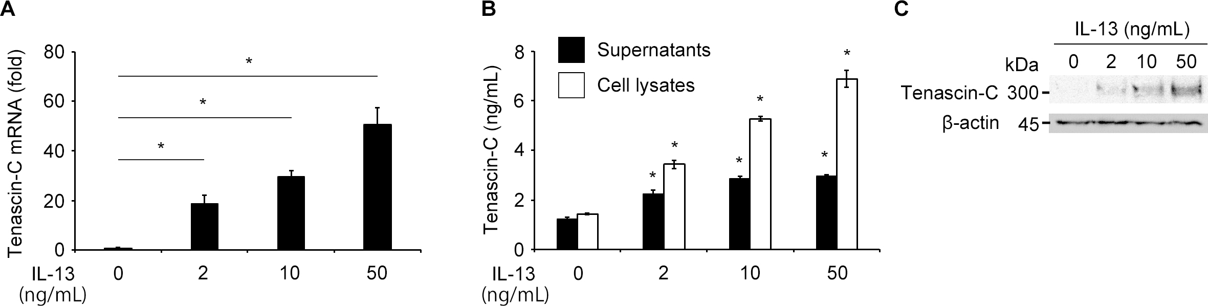
 Figure 3 of
Kobayashi, Mol Vis 2016; 22:436-445.
Figure 3 of
Kobayashi, Mol Vis 2016; 22:436-445.