Figure 4. Proliferative diabetic retinopathy epiretinal membranes. Immunohistochemical staining for CD45. A: Low power (original magnification 25X). B: High power (original magnification 40X). Double immunohistochemistry for CD45 (brown) and tissue factor pathway inhibitor
(TFPI; red) showing stromal cells and intravascular leukocytes (arrows) coexpressing CD45 and TFPI (C and D; original magnification 40X).
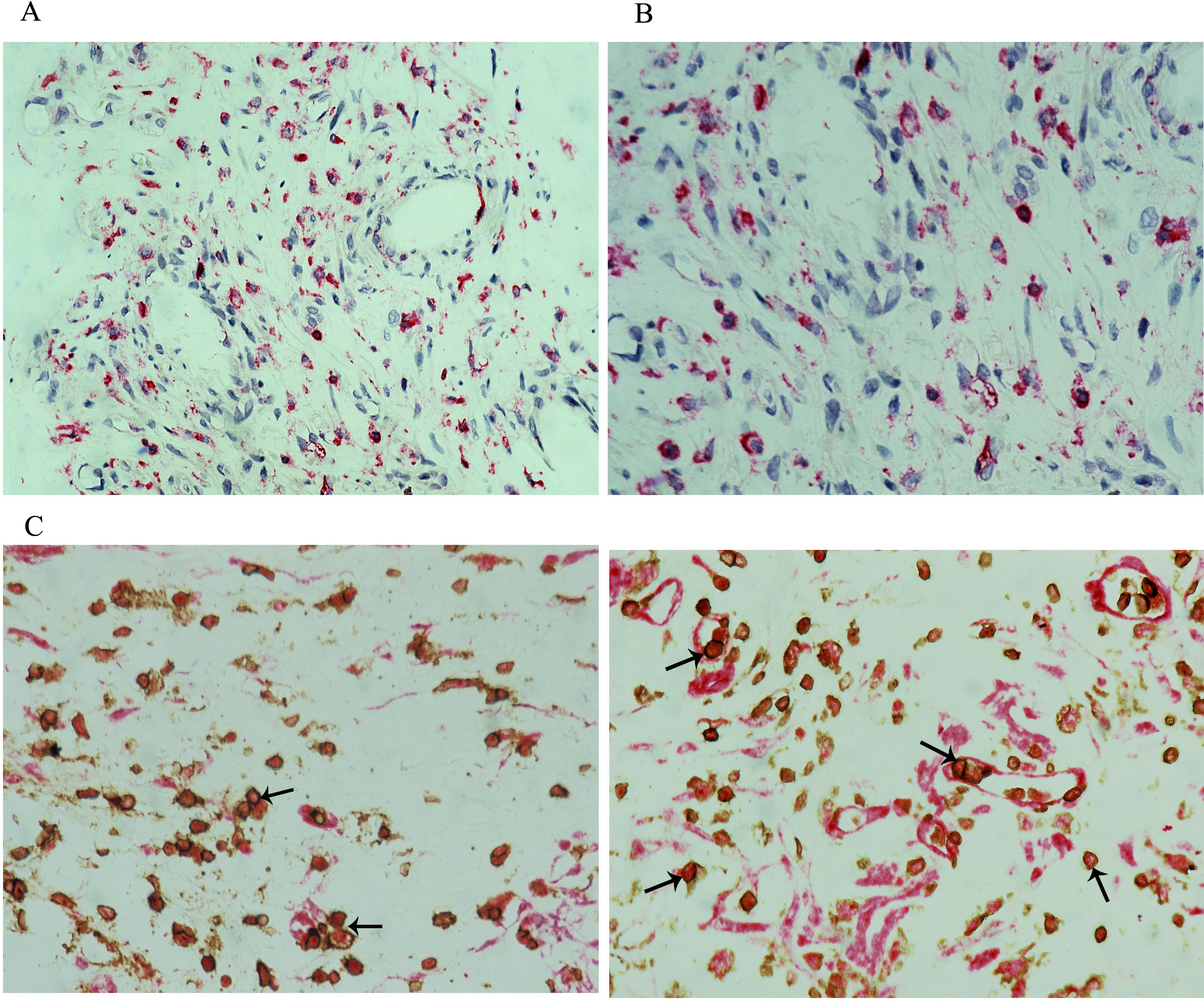
 Figure 4 of
Abu El-Asrar, Mol Vis 2016; 22:424-435.
Figure 4 of
Abu El-Asrar, Mol Vis 2016; 22:424-435.