Figure 3. Proliferative diabetic retinopathy epiretinal membranes. A: Immunohistochemical staining for tissue factor showing immunoreactivity in vascular endothelial cells (original magnification
40X). Immunohistochemical staining for tissue factor pathway inhibitor showing immunoreactivity in vascular endothelial cells
(arrows) and stromal cells (arrowheads). B: Low power (original magnification 25X). C: High power (original magnification 40X).
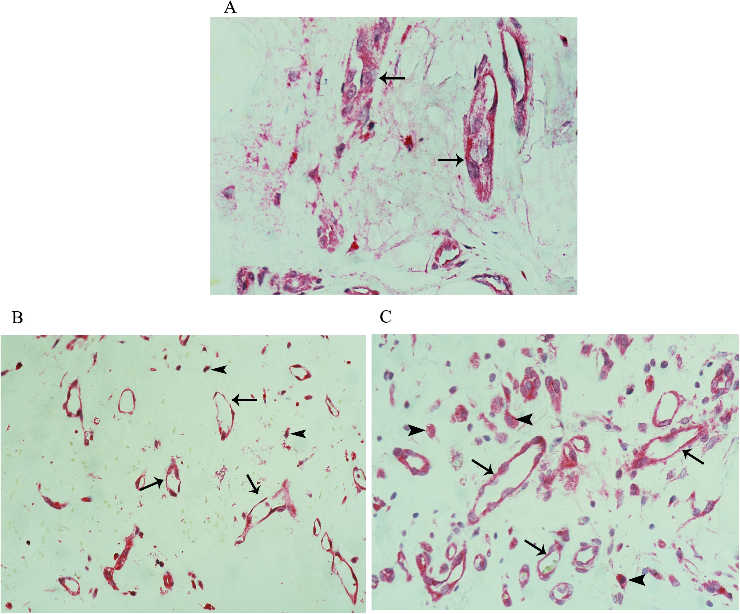
 Figure 3 of
Abu El-Asrar, Mol Vis 2016; 22:424-435.
Figure 3 of
Abu El-Asrar, Mol Vis 2016; 22:424-435.