Figure 2. Proliferative diabetic
retinopathy epiretinal membranes. A: A negative control
slide that was treated with an irrelevant antibody showed no
labeling (original magnification 40X). B:
Immunohistochemical staining for CD31 showing blood vessels
positive for CD31 (original magnification 40X).
Immunohistochemical staining for cathepsin L showing
immunoreactivity in vascular endothelial cells (arrows) and
stromal cells (arrowheads). C: Low power (original
magnification 25X). D: High power (original
magnification 40X).
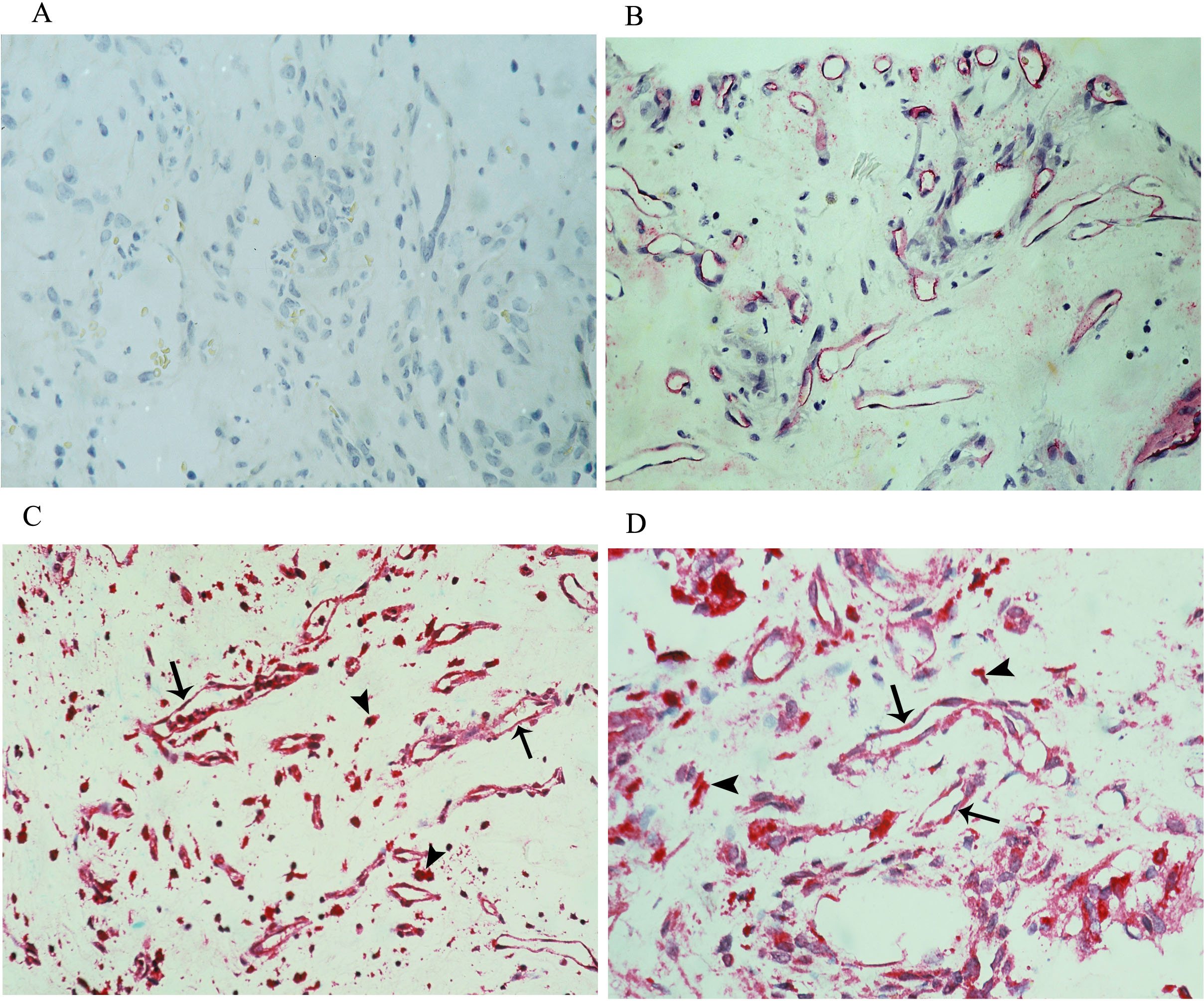
 Figure 2
of Abu El-Asrar, Mol Vis 2016; 22:424-435.
Figure 2
of Abu El-Asrar, Mol Vis 2016; 22:424-435.