Figure 10. In vitro study on Wistar rat lenses cultured for 24 h in Dulbecco’s modified Eagle’s medium: Immunoblots showing differential
staining intensity of some apoptotic cascade proteins in the three groups of lenses. Groups of lenses; Group I: Normal lenses
incubated in Dulbecco’s modified Eagle’s medium (DMEM) alone (control); Group II: Lenses incubated in DMEM that contained
sodium selenite (100 μM selenite/ml of DMEM; selenite-challenged, untreated); Group IIIa: Lenses incubated in DMEM and simultaneously
exposed to sodium selenite (100 μM selenite/ml of DMEM) and chrysin (200 μM chrysin/ml of DMEM; selenite-challenged simultaneously
chrysin-treated).
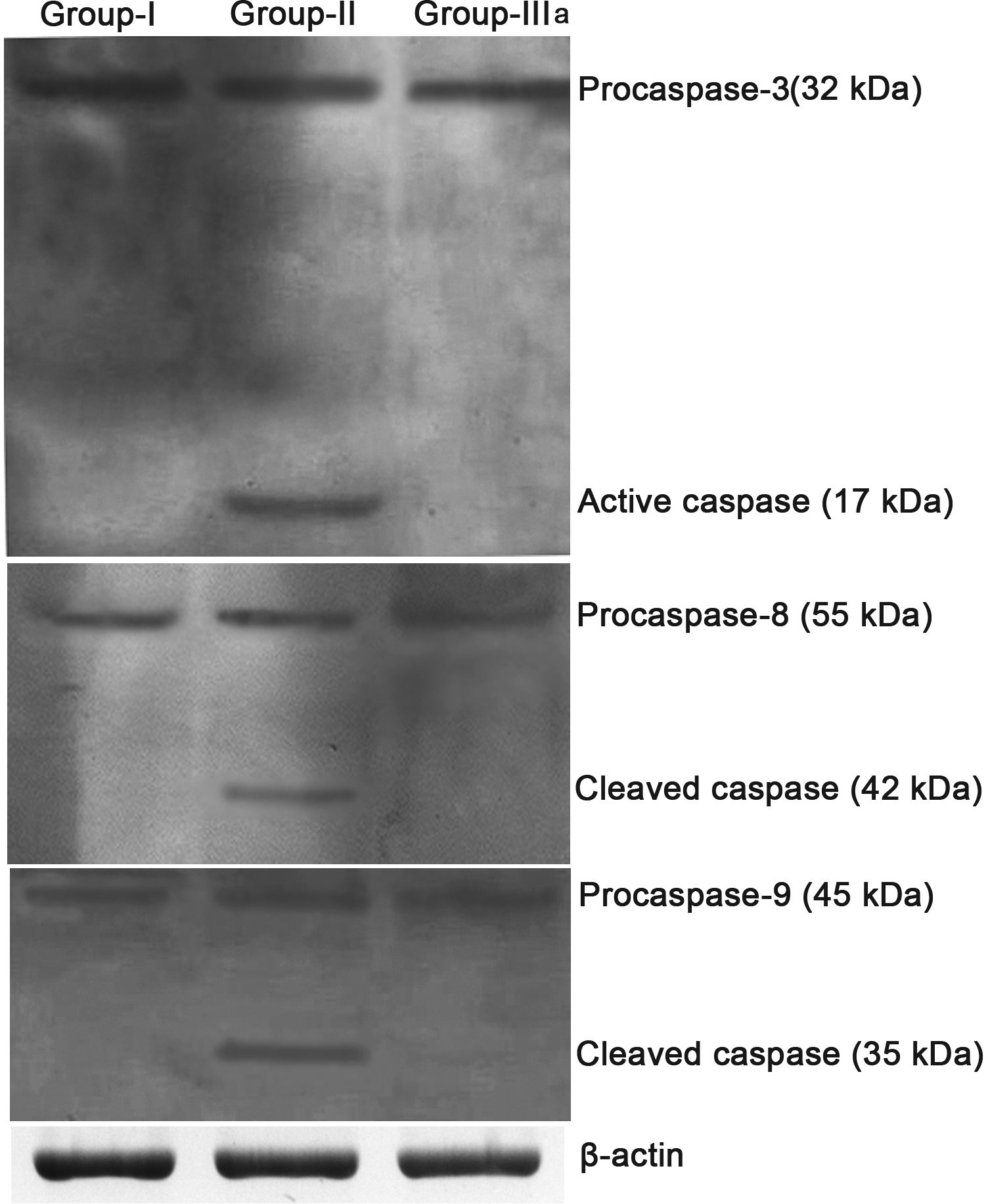
 Figure 10 of
Sundararajan, Mol Vis 2016; 22:401-423.
Figure 10 of
Sundararajan, Mol Vis 2016; 22:401-423.