Figure 7. RGC purity. Confocal double immunofluorescence of retinal ganglion cell (RGCs) from the two-step immunopanning (TIP) and two-step
immunopanning-magnetic (TIPM) methods shows the costaining of Thy-1 (A and E) and MAP2 (B and F). 4’,6-diamidino-2-phenylindole (DAPI) nuclear staining is shown in C and G, and in the merged images in (D) and (H). Cells negative for Thy-1 and MAP2 are regarded as non-RGCs and are marked with Arabic numbers (D and H). Visual counting of immunostained cells seeded at the same density demonstrates that seven cells in a 200X visual field
were not labeled by Thy-1 or MAP2 in the TIP groups while only three cells were negative in the TIPM group. Scale bars = 100
μm.
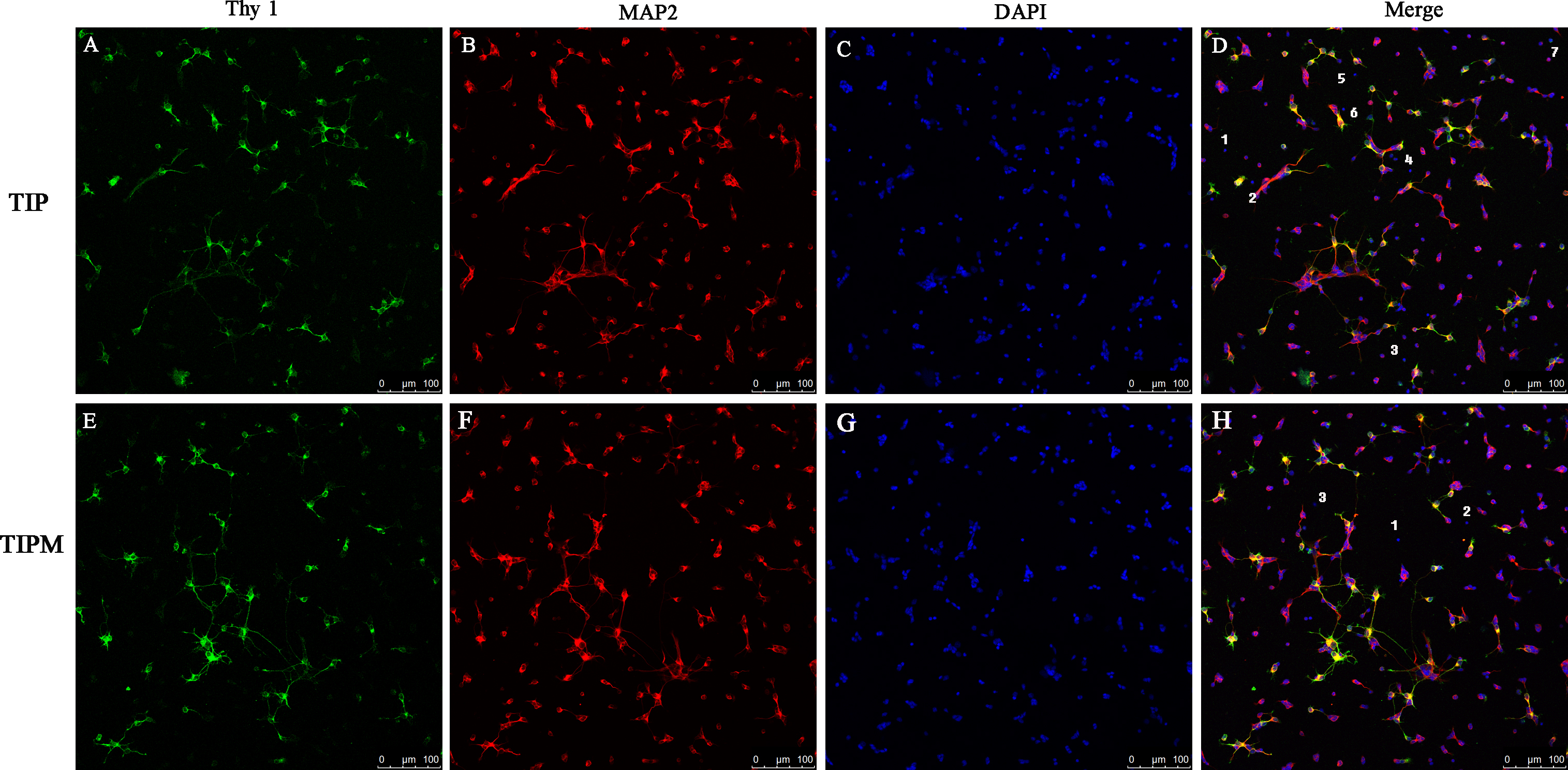
 Figure 7 of
Gao, Mol Vis 2016; 22:387-400.
Figure 7 of
Gao, Mol Vis 2016; 22:387-400.