Figure 5. RGC identification. Immunofluorescence of retinal ganglion cells (RGCs) at day 2 of culture shows costaining of γ-synuclein
(green) and MAP2 (red; A–D), TUJ1 (green), and Brn3b (red; E–H), Thy-1 (green), and Islet 1 (red; I–L). 4’,6-diamidino-2-phenylindole (DAPI) nuclear staining is shown in C, G, and K, and the merged images are shown in D, H and L. Scale bar = 50 μm.
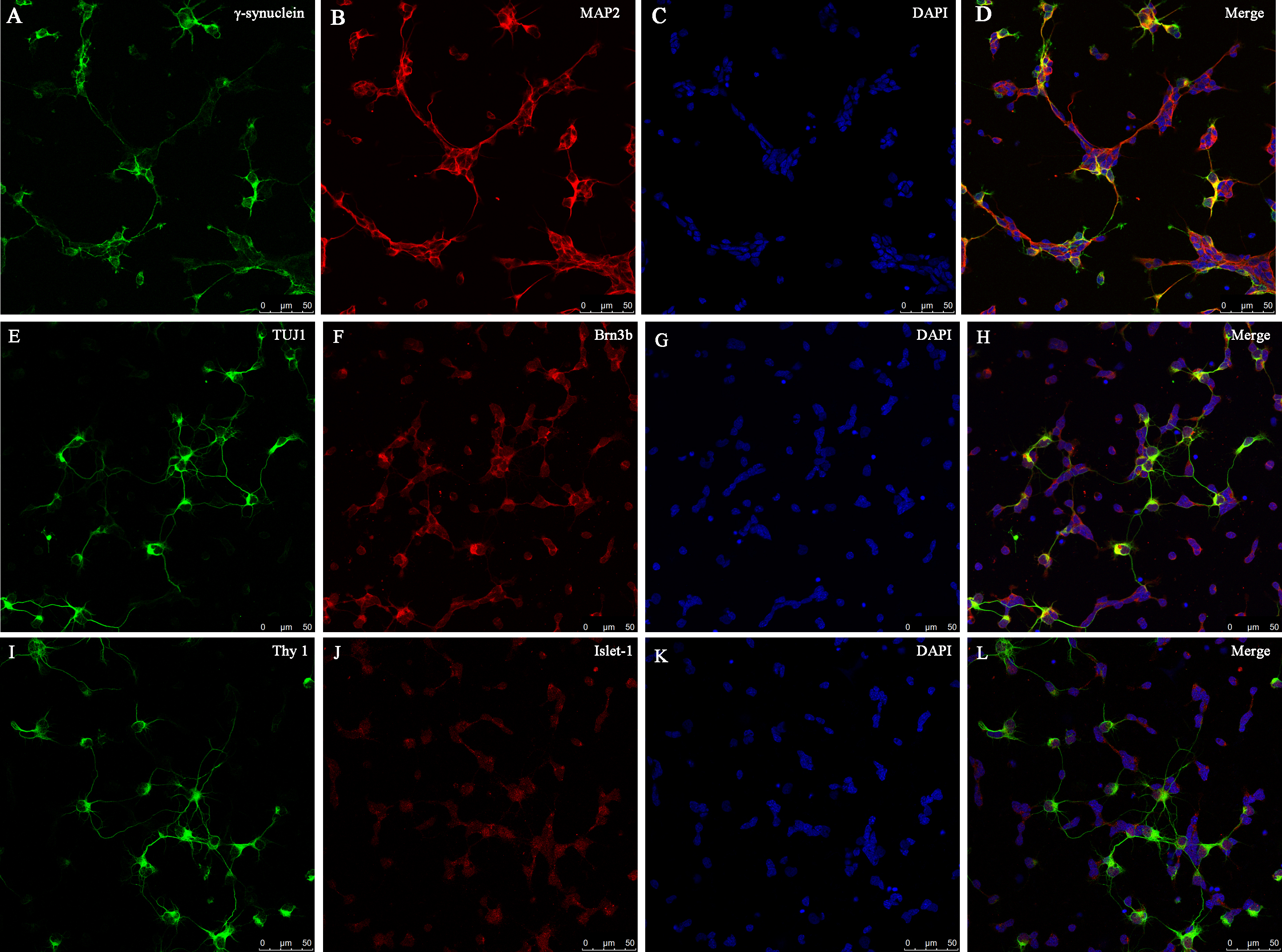
 Figure 5 of
Gao, Mol Vis 2016; 22:387-400.
Figure 5 of
Gao, Mol Vis 2016; 22:387-400.