Figure 2. Design of FC cell sorting gates. A: Blank control. Staining with control antibodies conjugated with phycoerythrin (PE). B: Retinal ganglion cell (RGC) sorting strategy. RGCs were identified by staining with PE-conjugated mAb directed against Thy-1
(R10 and R21). R10 is regarded as a low-purity group and R21 as a high-purity group. R16 consists mainly of non-RGCs. Y axes
= side scatter (SSC), X axes = forward scatter (FSC), fluorescence intensity on the FL2 channel.
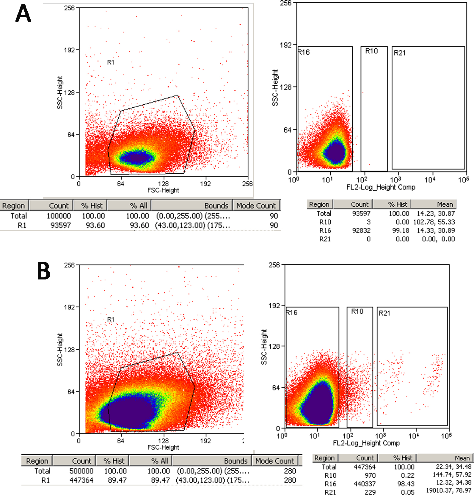
 Figure 2 of
Gao, Mol Vis 2016; 22:387-400.
Figure 2 of
Gao, Mol Vis 2016; 22:387-400.