Figure 5. HDGF stimulates retinal vascular leakage. A, B: Hepatoma-derived growth factor (HDGF) increases the permeability of human retinal microvascular endothelial cells (HRMVECs).
A: Schematic of in vitro permeability assay. HRMVECs were plated on the Transwell inserts and cultured until confluence. HDGF
(100 ng/ml), vascular endothelial growth factor (VEGF; 100 ng/ml), or PBS along with fluorescein isothiocyanate (FITC)–dextran
was added to the bottom chamber. B: Leaked FITC-dextran was quantified in the upper chamber at 6 h. C, D: Mice were intravitreally injected with HDGF (0.5 μg/eye), VEGF (0.2 µg/eye), or bovine serum albumin (BSA; 0.5 μg/eye) in
one eye with the contralateral eye for PBS. After 4 h, retinal vascular leakage was analyzed. C: Representative images of leaked mouse albumin detected with western blot using anti-mouse albumin. D: Retinal vascular leakage was quantified with the Evans blue assay. Leaked dye is normalized against the PBS-treated contralateral
eye. Mean ± standard error of the mean (SEM), n = 4 eyes/group, *p<0.05, **p<0.01, versus control or BSA, one-way ANOVA test.
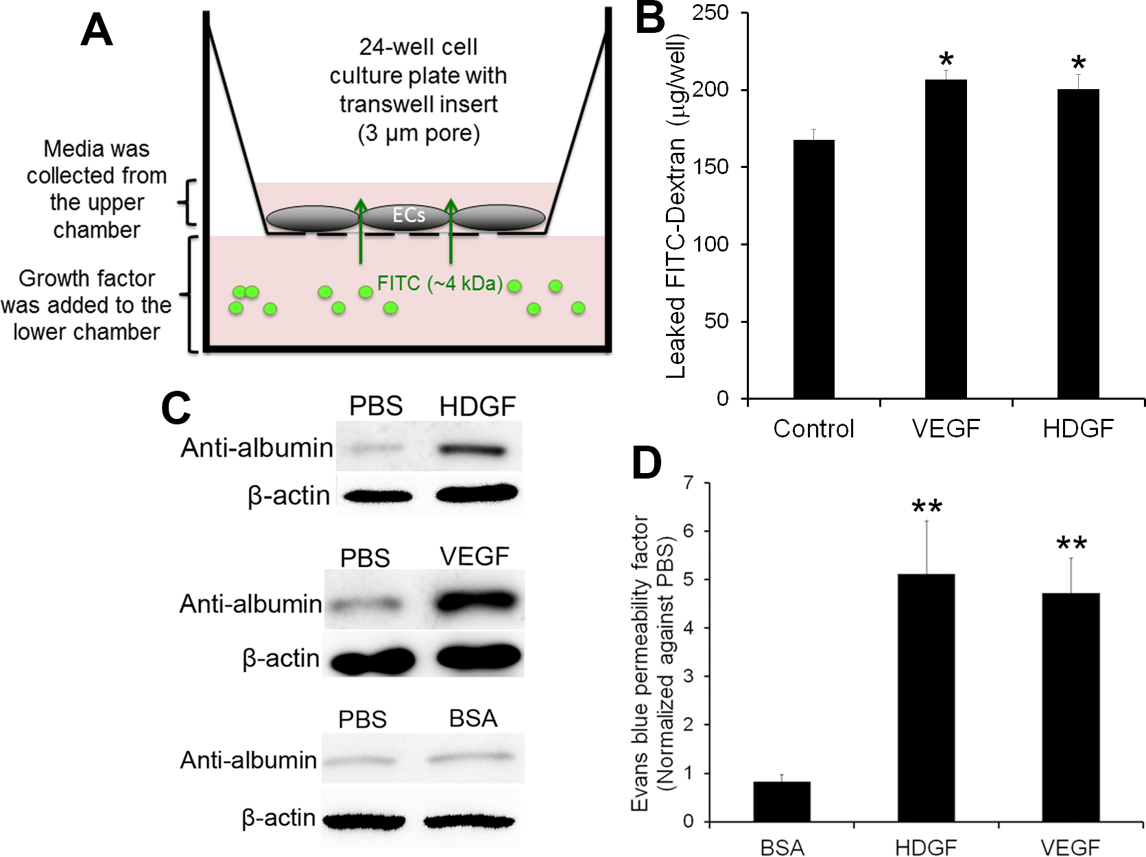
 Figure 5 of
LeBlanc, Mol Vis 2016; 22:374-386.
Figure 5 of
LeBlanc, Mol Vis 2016; 22:374-386.