Figure 4. HDGF induces corneal angiogenesis. A: Representative images of corneal angiogenesis assay. Small pieces of filter papers presoaked in hepatoma-derived growth
factor (HDGF; 1 µg/µl), vascular endothelial growth factor (VEGF; 100 ng/µl), or PBS were implanted in corneal pockets to
induce vascular sprouting for 6 days. * indicates filter paper position. B: Representative images of corneal blood vessels labeled with fluorescent DiI dye. C–E: Quantification of corneal angiogenesis assay. C: The number of blood vessels growing into the cornea. D: The number of branching points for corneal vessels. E: Comprehensive score for new vessels. Mean ± standard error of the mean (SEM), n=5 eyes/group, **p<0.01, ***p<0.001, ****p<0.0001,
versus PBS, one-way ANOVA test.
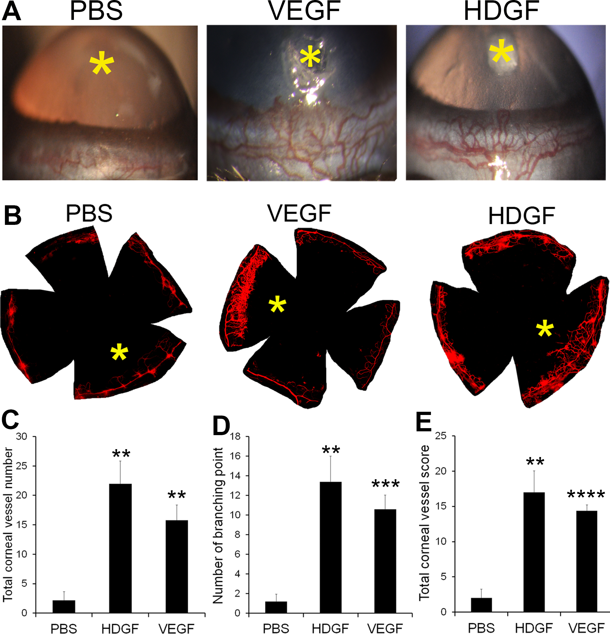
 Figure 4 of
LeBlanc, Mol Vis 2016; 22:374-386.
Figure 4 of
LeBlanc, Mol Vis 2016; 22:374-386.