Figure 3. HDGF stimulates endothelial migration. A: Representative images of endothelial migration by wound healing assay. Human retinal microvascular endothelial cells (HRMVECs)
were cultured in 12-well plates to confluence. A scratch was created in each well. Hepatoma-derived growth factor (HDGF; 500
ng/ml), vascular endothelial growth factor (VEGF; 100 ng/ml), or PBS was incubated with the cells for 20 h. Bar = 100 μm.
B: The percentage of the denuded area covered by migrated cells within the original scratch was quantified. This assay was
independently performed three times with similar outcomes. Mean ± standard error of the mean (SME), n = 3 wells/group, *p<0.05,
versus control, one-way ANOVA test.
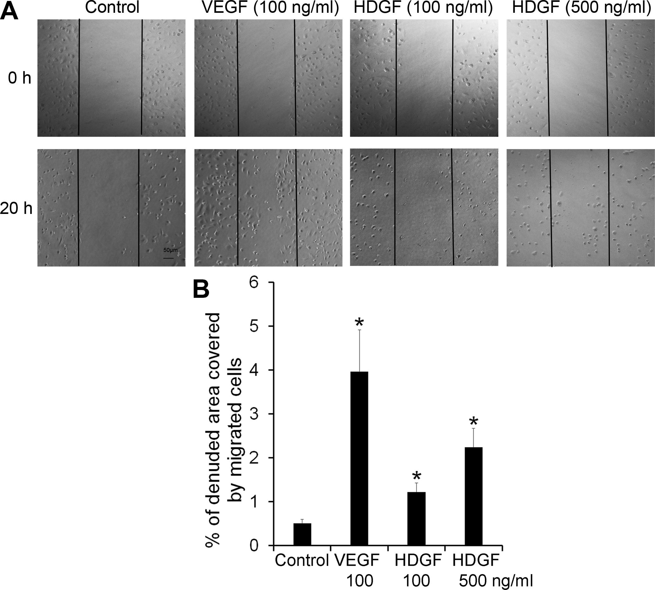
 Figure 3 of
LeBlanc, Mol Vis 2016; 22:374-386.
Figure 3 of
LeBlanc, Mol Vis 2016; 22:374-386.