Figure 1. HDGF expression in the retina. A: Hepatoma-derived growth factor (HDGF) expression in the mouse retina was detected with reverse transcription PCR (RT–PCR).
GAPDH was included as a positive control. B: Western blot analysis of HDGF expression in the mouse retina. Mouse total brain homogenate (middle lane) and purified recombinant
HDGF (left lane) with extra C-terminal polyhistidine were included as positive controls. Five hundred µg protein/lane for
retinal or brain homogenate. One µg protein/lane for purified HDGF. The predicted molecular weight (MW) for HDGF is 25.5 kDa.
C: Immunohistochemical analysis of HDGF expression in the mouse retina. HDGF is predominantly expressed in the inner and outer
nuclear layers, photoreceptor outer segments, and choroid. HDGF was also detected in the retinal ganglion cell layer and the
photoreceptor inner segments at relatively low levels. Few HDGF signals were found in the inner and outer plexiform layers.
Bar = 100 μm. D: HDGF is present in the cytoplasmic and nuclear fractionations by western blot. These results were validated three times
with similar outcomes.
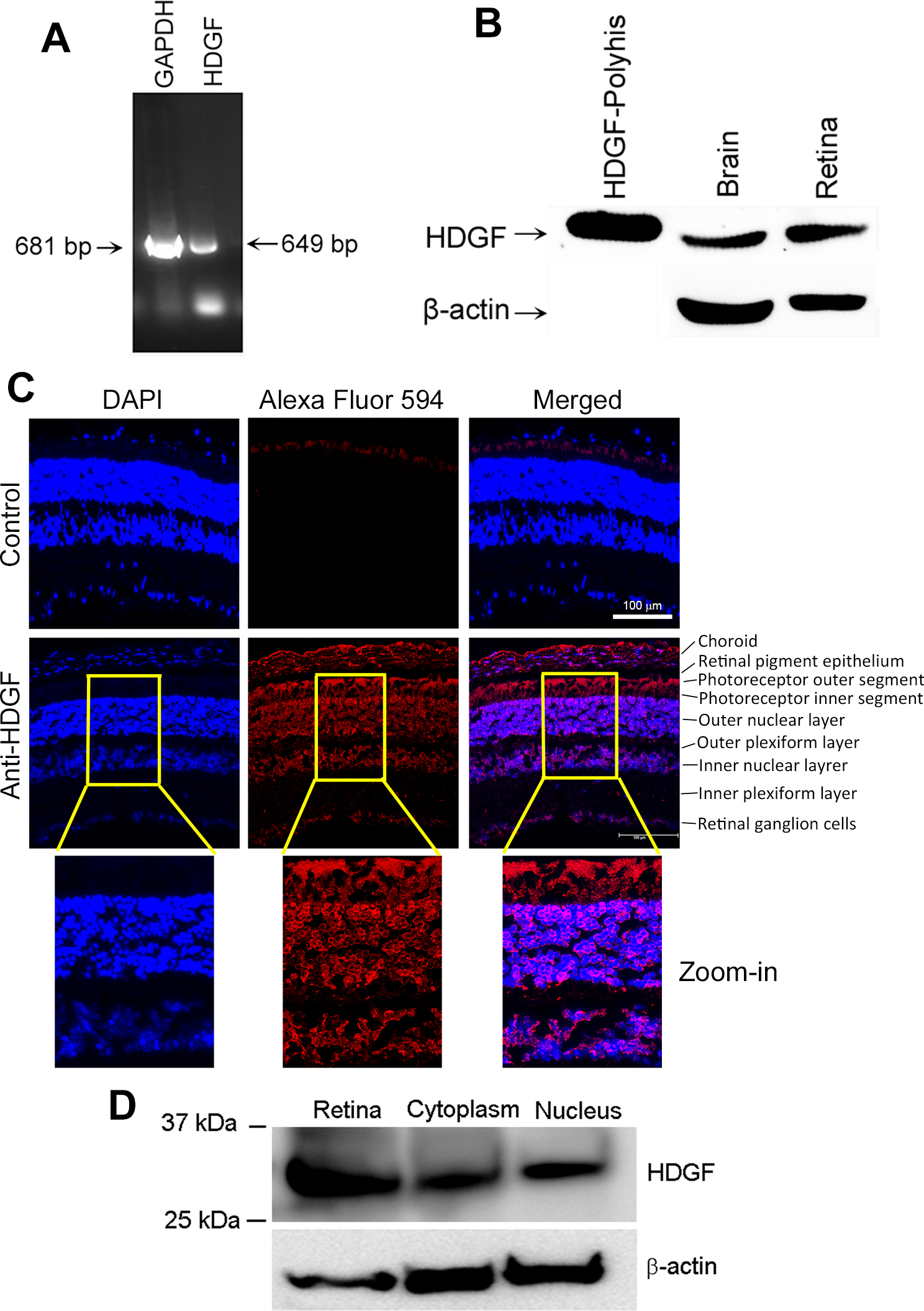
 Figure 1 of
LeBlanc, Mol Vis 2016; 22:374-386.
Figure 1 of
LeBlanc, Mol Vis 2016; 22:374-386.