Figure 4. Phenotype. Patient II-1 with autosomal dominant retinitis pigmentosa (adRP) and a de novo mutations in RHO135 (p.R135W), showing normal fundi, a slightly reduced optical coherence tomography (OCT) scan and a reduced multifocal electroretinography
(mfERG). This patients with normal full-field electroretinography (ffERG) and visual fields shows the importance of assessing
central retinal structure and function in the diagnostics of RP.
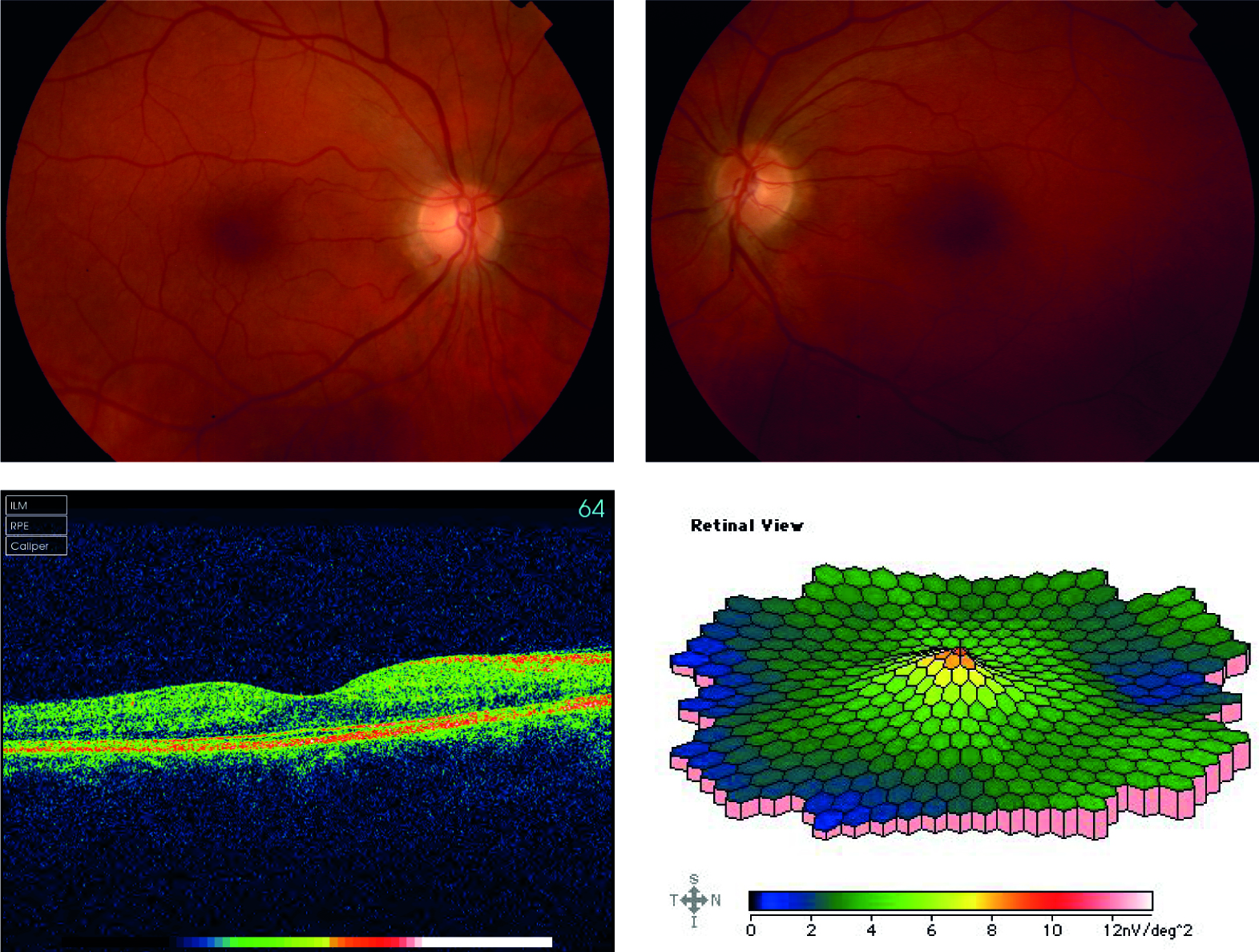
 Figure 4 of
Abdulridha-Aboud, Mol Vis 2016; 22:362-373.
Figure 4 of
Abdulridha-Aboud, Mol Vis 2016; 22:362-373.