Figure 3. Phenotype. The top figure shows fundus photos, optical coherence tomography (OCT) and multifocal electroretinography (mfERG)
results for the family with the PRPF31 (p.IVS6+1G>T) mutation, demonstrating affected patients from three generations and increasing retinal pathology with increasing
age. The bottom figure shows fundus photos, OCT, and mfERG results for the family with RHO135 (p.R135W) mutations, demonstrating marked intrafamilial variability in the clinical phenotype, regarding retinal structure
and function. For patient III-2, the mfERG is from her left eye.
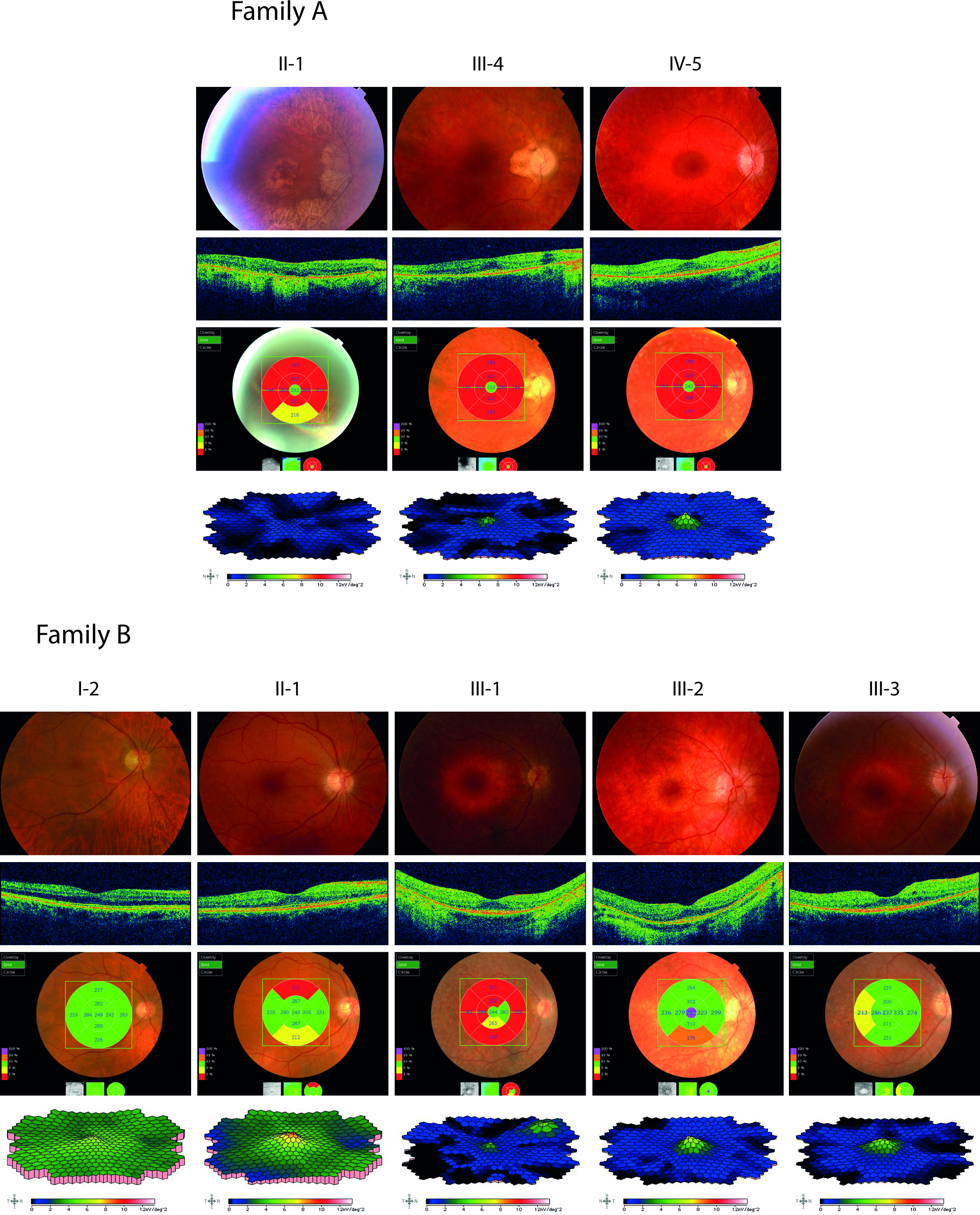
 Figure 3 of
Abdulridha-Aboud, Mol Vis 2016; 22:362-373.
Figure 3 of
Abdulridha-Aboud, Mol Vis 2016; 22:362-373.