Figure 1. Experimental design. Cultured, myelin-specific Th1 (interleukin (IL)-12-treated) or Th17 (IL-23-treated) cells were injected
into 15 naïve recipient mice each, and optokinetic reflexes (OKTs) and disease severity were assessed daily. At approximately
1, 2, and 3 weeks post-induction, five mice from each group were harvested. The optic nerves were histologically assessed
for inflammation, and retinal ganglion cells (RGCs) were counted in retinal flatmounts.
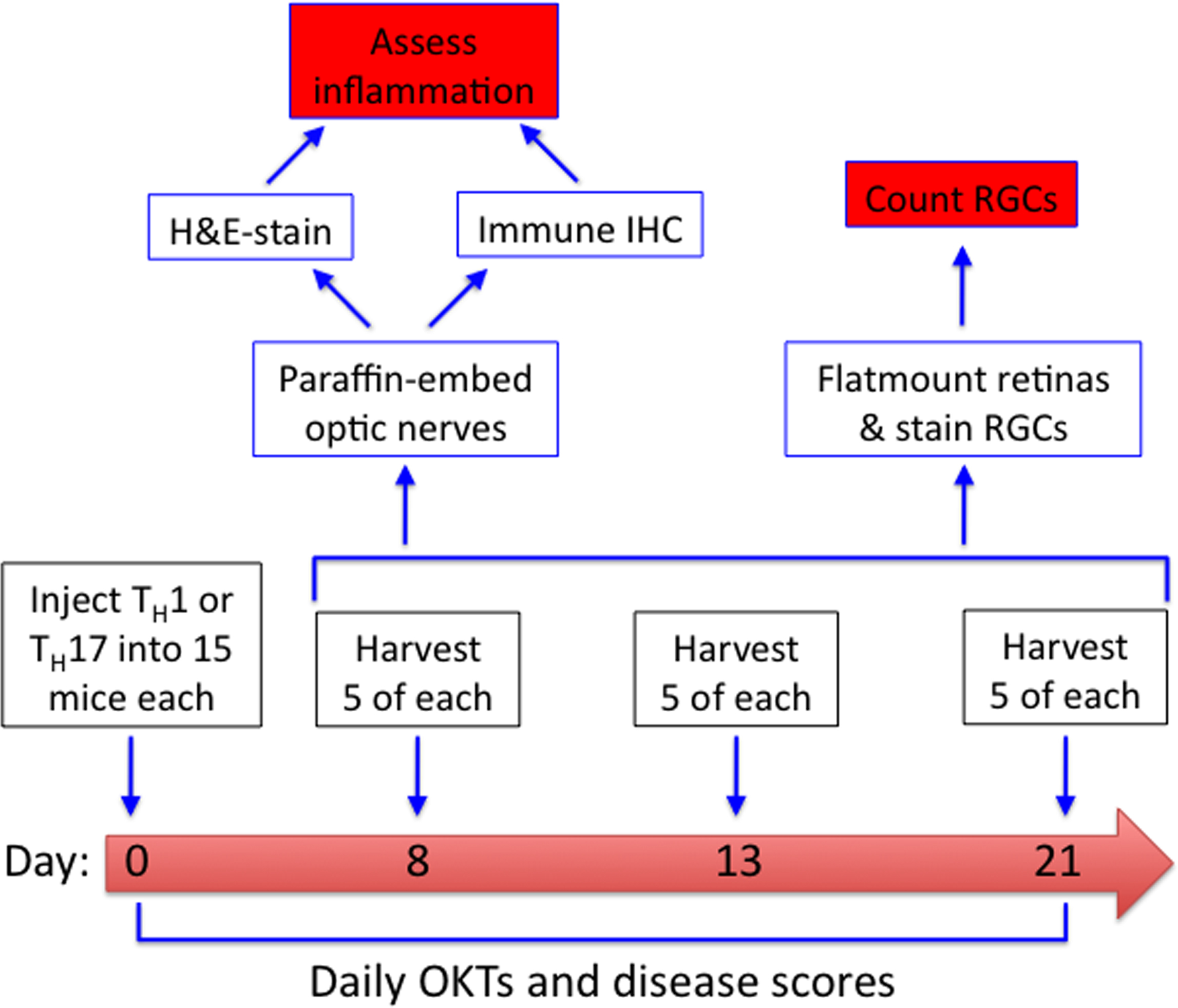
 Figure 1 of
Larabee, Mol Vis 2016; 22:332-341.
Figure 1 of
Larabee, Mol Vis 2016; 22:332-341.