Figure 3. Immunolocalization of corneal endothelial cell lateral membrane markers by widefield fluorescence microscopy. A: Reaction of intact tissues with ZO-1 antibody highlights the regular polygonal outlines of cells seen at the level of tight
junctions. B: Staining for the cell adhesion protein neural cell adhesion molecule (NCAM), however, reveals that much of the interacting
surface is in the form of a complex arrangement of membrane folds. When viewing whole cells, this gives the impression of
extensive membrane ruffling.
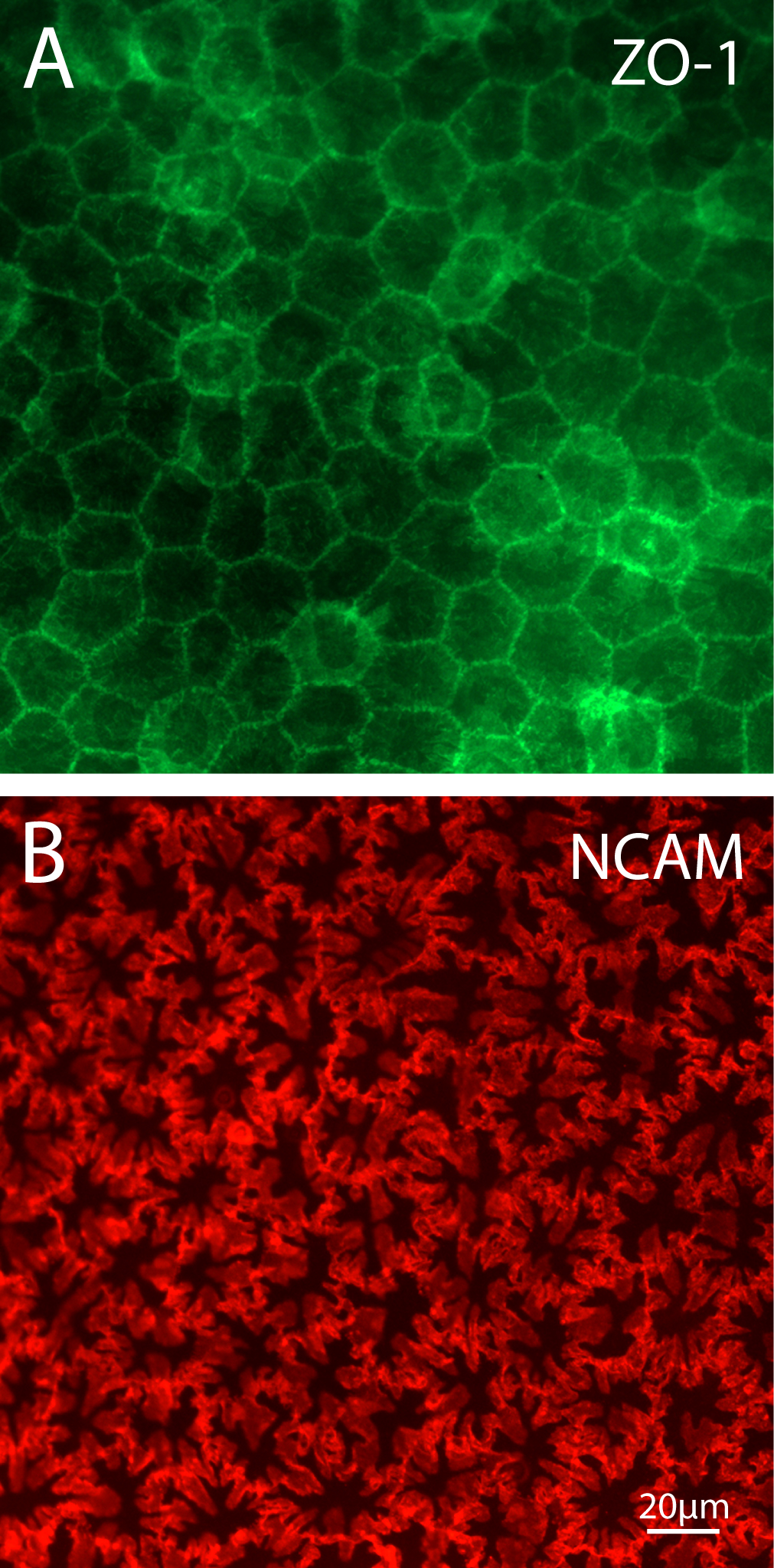
 Figure 3 of
Harrison, Mol Vis 2016; 22:31-39.
Figure 3 of
Harrison, Mol Vis 2016; 22:31-39.