Figure 2. Gallery of endothelial cell surface representations obtained following 3D reconstruction from confocal image stacks. A–F: Viewed from their anterior surface at 3/4 perspective, mosaic analysis with double markers (MADM)-labeled corneal endothelial
cells present flat apical membranes in the form of a plateau region (indicated by asterisks in B, D, and F) that overlaps and partially obscures the dendritic lateral processes. In some cases, the processes can be seen to taper
as they extend peripherally and ultimately give rise to smaller branches (arrows in A and C).
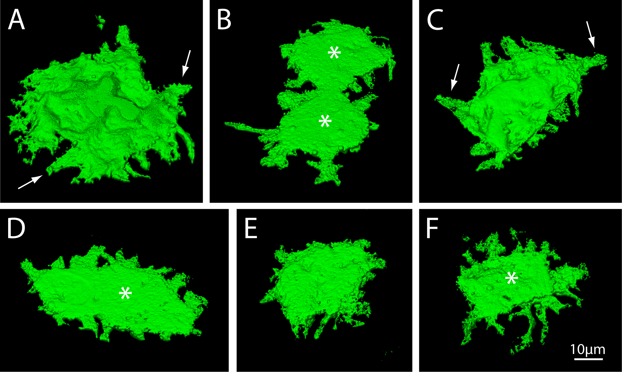
 Figure 2 of
Harrison, Mol Vis 2016; 22:31-39.
Figure 2 of
Harrison, Mol Vis 2016; 22:31-39.