Figure 8. No difference in scotopic ERG response with drug treatment. Dark-adapted electroretinogram (ERG) amplitudes measured at three
flash intensities for mice treated with 0 mg/kg (control, n = 12), 0.5 mg/kg (low dose, n = 15), or 3.0 mg/kg (high dose,
n = 12) of xaliproden for 4 months. A: a-wave amplitudes at 2 months of treatment. B: a-wave amplitudes at 4 months of treatment. C: b-wave amplitudes at 2 months of treatment. D: b-wave amplitudes at 4 months of treatment. Scotopic ERGs were elicited with 1 msec flashes of white light at 0 dB (2.68
cds/m2), −10 dB (0.18 cds/m2), and −20 dB (0.02 cds/m2). (n = 12). With the exception of the b-wave response at 2 weeks at the −10 dB flash intensity (p<0.05), we observed no statistically
significant difference among the three treatment groups. E: Following 2 min of white light exposure, the photopic b-wave ERG amplitudes at −10 dB (0.18 cds/m2) were measured following 2 months and 4 months of treatment with xaliproden. Control = Vehicle, low dose = 0.5 mg/kg, high
dose = 3.0 mg/kg. n = 12; (*p<0.05, high dose versus low dose; high dose versus control not significant.) Error bars are standard
error of the mean.
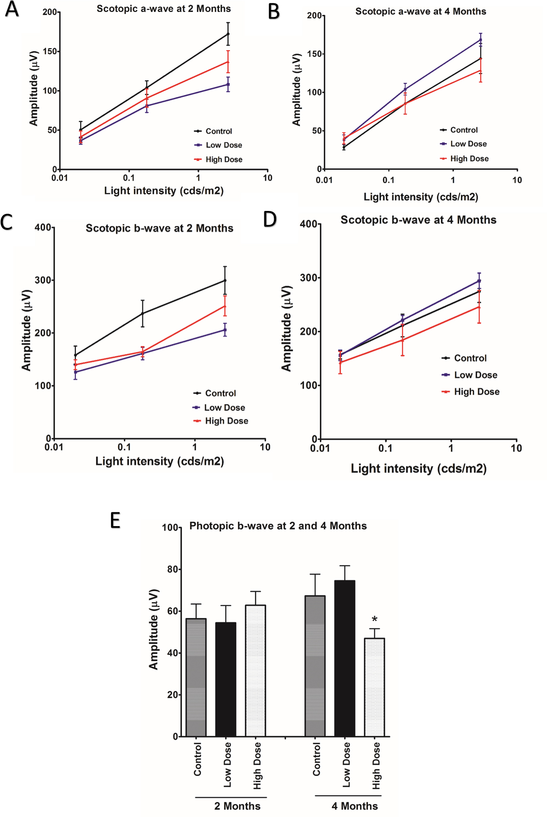
 Figure 8 of
Ahmed, Mol Vis 2016; 22:294-310.
Figure 8 of
Ahmed, Mol Vis 2016; 22:294-310.