Figure 3. Anti-cytokeratin-10 immunohistochemistry. Representative immunostaining images of sections from the cornea stained with anti-cytokeratin-10
(Green). Nuclei were stained with 4′,6-diamidino-2-phenylindole (DAPI), and sections were viewed under a confocal microscope
(Original magnification: 630X). A: The control group. B: The dry eye (DE) group. C: The 50 mg/kg ethanol extract of Diospyros kaki (EEDK)-treated group. D: The 10 mg/kg EEDK-treated group. E: The positive-control group (Refresh Plus, preservative-free eye drops, Allergan). The results shown are representative of
three independent experiments (n = 8 mice per group).
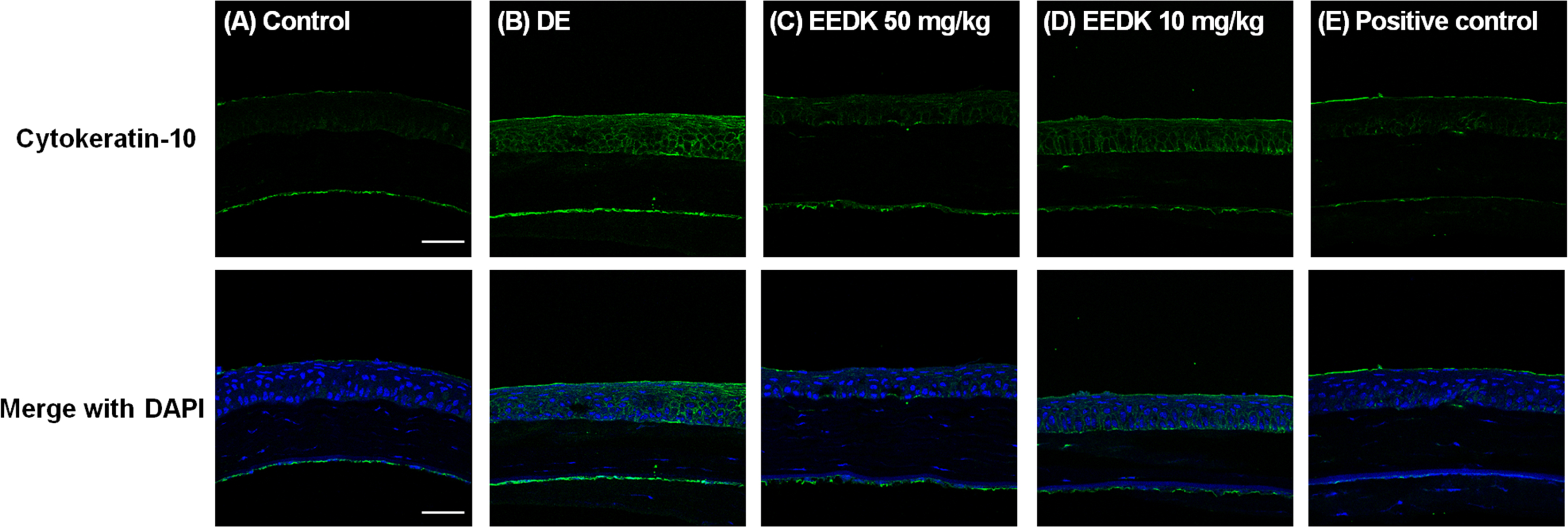
 Figure 3 of
Kim, Mol Vis 2016; 22:284-293.
Figure 3 of
Kim, Mol Vis 2016; 22:284-293.