Figure 1. Mouse retinal Müller cells express the canonical circadian clock genes. A: 4’,6-diamidino-2-phe-nylindole (DAPI) staining showing all cell nuclei (blue) in a fluorescence photo-micrograph of a purified
Müller cell culture. B: Glutamine synthase (GS) antibody staining showing Müller cells (green). C: Merged image of A and B, showing the high degree of purity of the Müller cell culture. D: Clock gene expression from a purified Müller cells culture with quantitative PCR. Nitric oxide synthase (NOS), which is
present in many retinal neurons but not in Müller cells, was used as a negative control for the purity of the cultures.
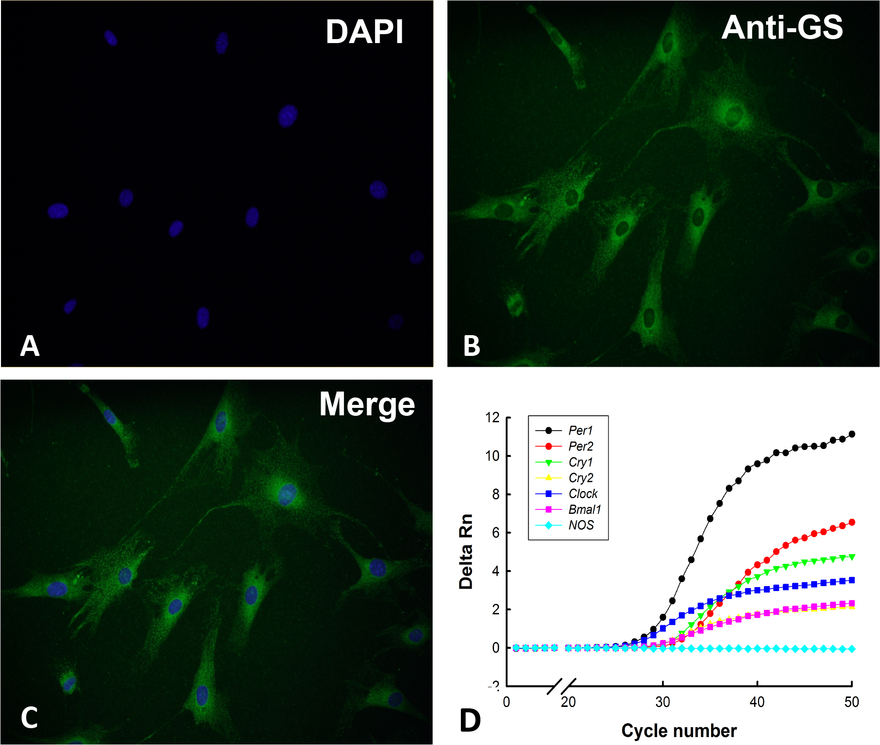
 Figure 1 of
Xu, Mol Vis 2016; 22:275-283.
Figure 1 of
Xu, Mol Vis 2016; 22:275-283.