Figure 3. Norgestrel potentiates increases in LIF in degenerating rd10 retinas. A: Relative expression of Lif in P15 and P20 rd10 retinas compared to age-matched wild-type C57BL/6 controls, as measured with real-time (RT) PCR, with
fold change compared to the geometric mean of three endogenous reference genes. Error bars denote
±
standard error of the mean (SEM). B and C: Immunostaining of LIF (green) and glutamine synthetase (GS; red) in rd10 retinas at P15 (B) and P20 (C). Increased LIF levels are observed in mice that received the norgestrel diet (lower panels) during retinal degeneration,
before photoreceptor loss (P15) and during photoreceptor loss (P20). Colocalization (yellow staining) is observed with a sub-set
of Müller glial cells in the OPL and at the Müller glia end feet in the inner plexiform layer (IPL). Scale bars represents
50 µm. Results are representative of n = 3 mice.
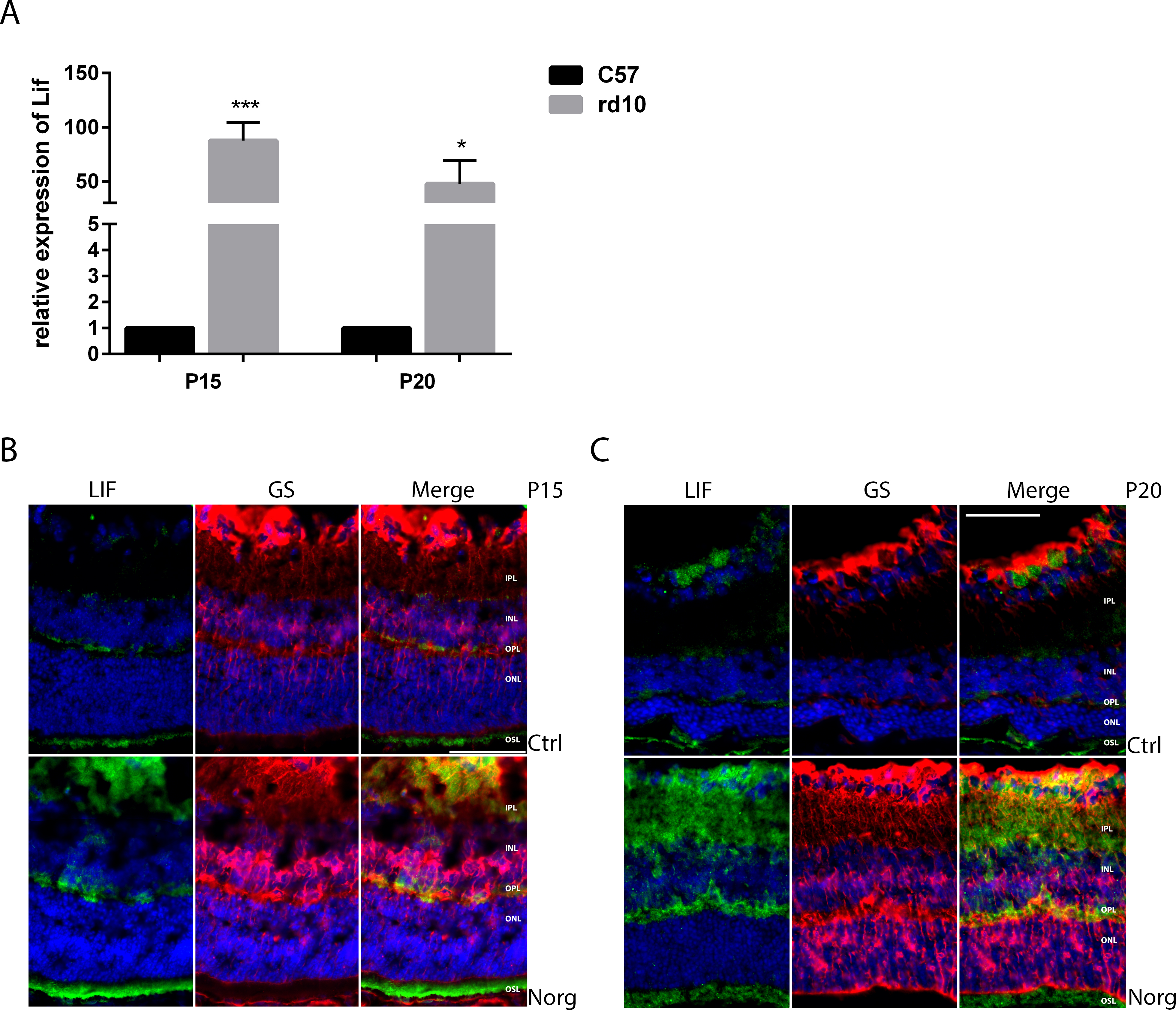
 Figure 3 of
Byrne, Mol Vis 2016; 22:264-274.
Figure 3 of
Byrne, Mol Vis 2016; 22:264-274.