Figure 1. Expression of TrkA in Müller cells. Primary mouse Müller cells were identified with immunofluorescence staining of glutamine
synthetase (GS; red), a specific marker of Müller cells. TrkA (green) was expressed abundantly in the mouse Müller cells.
Scale bar=50 μm.
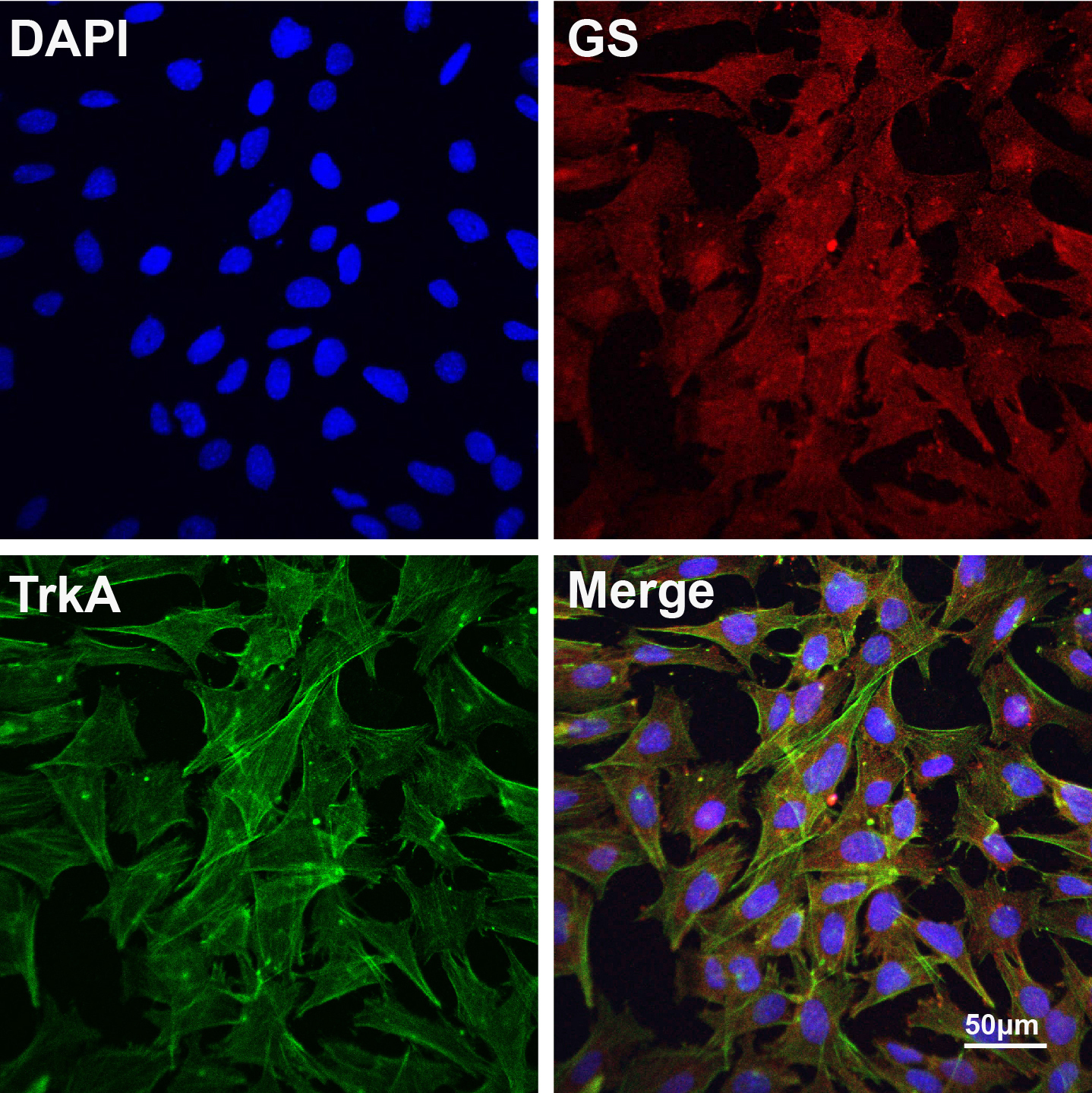
 Figure 1 of
Wang, Mol Vis 2016; 22:254-263.
Figure 1 of
Wang, Mol Vis 2016; 22:254-263.