Figure 5. Neuroretina immunohistochemistry during the culture and coculture period. In the freshly isolated retina explants (A), RHO immunoreactivity (green) showed the normal appearance of rod outer segments (OS). At 9 days of neuroretina explants
culture alone (B), RHO was scarcely detected, and the inner segments (IS) were not discernable. Photoreceptor nuclei appeared at the IS region
(B, arrowhead). At 9 days in coculture with RPE cells (C), RHO was displaced to the short and swollen rod IS and to the outer nuclear layer (arrows). In the freshly isolated retina
explants (D), CB (red) showed cone photoreceptors with normal morphology. At 9 days of neuroretina culture alone (E), cone morphology was markedly altered as revealed with CB immunostaining. At 9 days in coculture with the RPE (F), cone morphology underwent degeneration. In the freshly isolated retina explants (G), CRALBP (green) showed Müller cells with normal morphology. Reduced immunostaining for GFAP (red) was observed. At 9 days
of neuroretina culture alone (H), GFAP was upregulated at the cytoplasm of glial cells, and GFAP-positive extensions formed a layered-like structure outside
the retinal tissue (arrows). At 9 days in coculture with RPE (I), CRALBP was scarcely detected, and GFAP was reduced compared to explants cultured alone. GFAP-positive processes were present
over the outer limiting membrane (arrows). OS=outer segments; IS=inner segments; ONL=outer nuclear layer; INL=inner nuclear
layer; GCL=ganglion cell layer. Scale bars=10 μm.
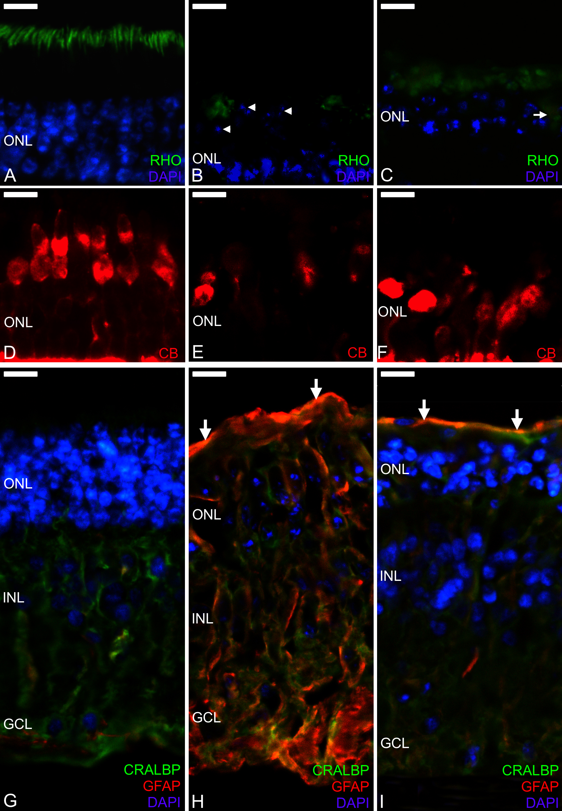
 Figure 5 of
Di Lauro, Mol Vis 2016; 22:243-253.
Figure 5 of
Di Lauro, Mol Vis 2016; 22:243-253.