Figure 3. Cellular morphology and immunochemistry of RPE cells during the culture and coculture period. At day 4 of culture in the flasks
(A), the RPE cells formed clusters of pigmented, polygonal cells. At day 7 in the flasks (B), the RPE cells reached confluence and maintained morphological characteristics. Dedifferentiated cells were present (B, arrowheads). At 9 days of coculture with the neuroretina explants (C), the RPE cells in the cell culture inserts were confluent, monolayered, pigmented, and polygonal-shaped (C, insert). Dedifferentiated cells were still present (C, arrowheads). At 9 days of coculture, the RPE cells in the cell culture inserts showed variable ZO-1 immunoexpression at
the cell periphery (D, green; nuclei, blue) and maintained cytoplasmic RPE65 (E, red; nuclei, blue). CRALBP was not detectable (F) Scale bars=100 μm (A–C) and 20 μm (D–F).
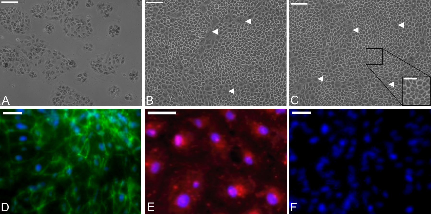
 Figure 3 of
Di Lauro, Mol Vis 2016; 22:243-253.
Figure 3 of
Di Lauro, Mol Vis 2016; 22:243-253.