Figure 1 of
Di Lauro, Mol Vis 2016; 22:243-253.
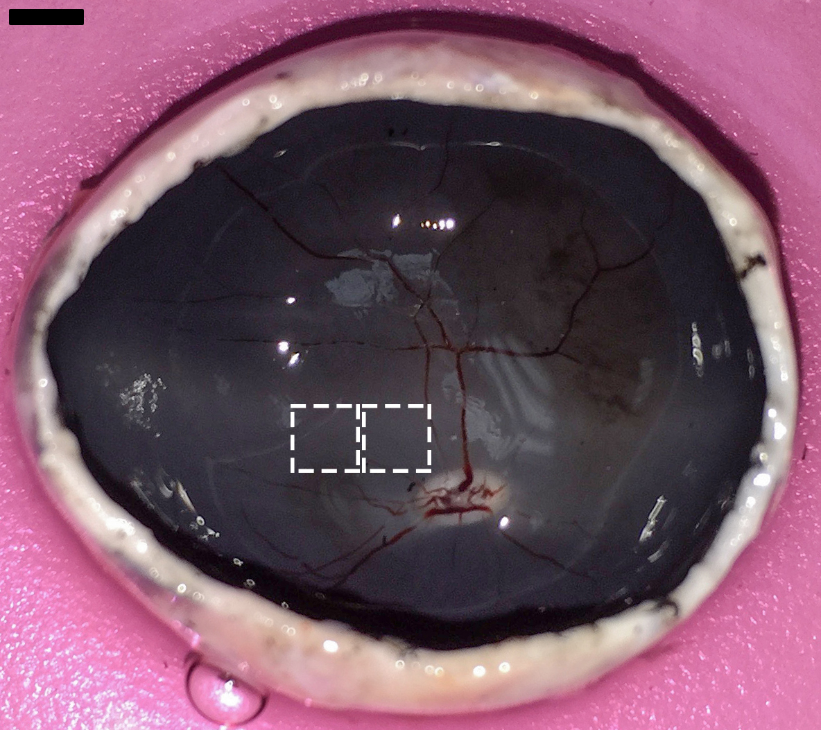
Figure 1.
Neuroretina explant tissue sampling in porcine globe. Two neuroretina explants (5×5 mm) were obtained from each eye at the porcine cone-enriched visual streak superotemporal to the optic disc. Scale bar=5 mm.
 Figure 1 of
Di Lauro, Mol Vis 2016; 22:243-253.
Figure 1 of
Di Lauro, Mol Vis 2016; 22:243-253.