Figure 2. Fundus photography and OCT test of normal and affected member of this pedigree. Fundus photograph of the normal individual
III:10 (A) and the patient II:3 (B) revealed that the patients had typical RP symptoms including attenuated retinal arterioles, peripheral intraretinal pigment
deposits in a bone-spicule configuration, degenerated macula and diffused mottling of retinal pigmented epithelium. Optical
coherence tomography (OCT) images of the normal individual III:10 (C) and the patient II:3 (D) showed that the patient had marked thinning and disruption of the photoreceptor layer and the retinitis pigment epithelium.
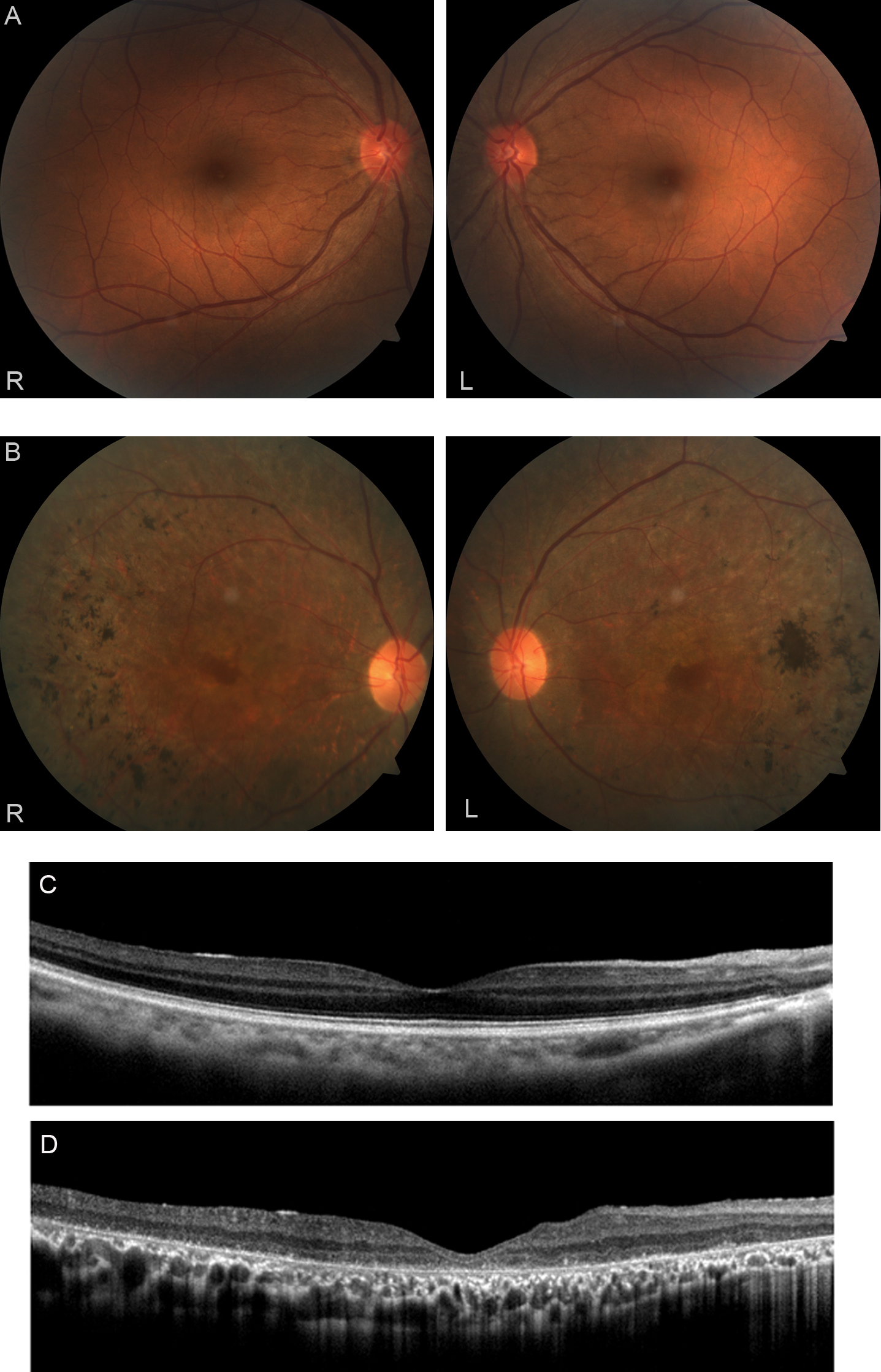
 Figure 2 of
Gao, Mol Vis 2016; 22:234-242.
Figure 2 of
Gao, Mol Vis 2016; 22:234-242.