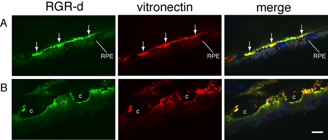
Figure 8. Association of extracellular RGR-d and vitronectin in Bruch’s membrane and basal deposits. The RPE-choroid section from a
78-year-old male (same donor as in
Figure 6) was incubated first with DE21 antibody and anti-rabbit IgG conjugated to FITC and subsequently with anti-vitronectin monoclonal
antibody and anti-mouse IgG antibody conjugated to Cy3.
A: Confocal image plane reveals immunoreactive basal deposits (arrows).
B: Different confocal plane of the same field as in top panels with images that exhibit immunoreactivity in the choroid and
intercapillary regions of Bruch’s membrane. RGR-d immunoreactivity is detected in the basal deposits and distributed among
aggregates or speckles in Bruch’s membrane. Specific immunolabeling of vitronectin was also observed in each confocal image.
The merged images with DAPI counterstain indicate significant co-localization of RGR-d and vitronectin in basal deposits and
Bruch’s membrane. Autofluorescence is visible in RPE cells. C, choroidal capillary. Scale bar, 10 μm.
 Figure 8 of
Kochounian, Mol Vis 2016; 22:213-223.
Figure 8 of
Kochounian, Mol Vis 2016; 22:213-223.