Figure 5. Differences in amount and targeting of RGR-d in the RPE and Bruch’s membrane of young and old donors. All sections were from
unfixed tissue and stained in parallel by immunohistochemical (left panels) and immunofluorescent (right panels) labeling
of RGR-d with the DE21 antibody. A: RGR-d labeling in an 87-year-old female donor is predominantly in Bruch’s membrane and the choriocapillaris layer but weak
or absent in RPE cells. B: Relatively high amounts of RGR-d are present in the RPE of a young 20-year-old female donor, as indicated by strong immunohistochemical
staining. Intense immunofluorescent labeling was present in the basolateral plasma membrane (arrowheads) in most RPE cells
in the young donor. C: Negative control labeling of 20-year-old donor tissue. Scale bar, 40 μm.
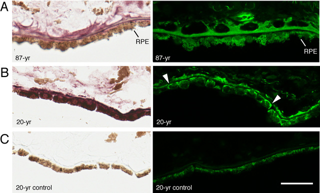
 Figure 5 of
Kochounian, Mol Vis 2016; 22:213-223.
Figure 5 of
Kochounian, Mol Vis 2016; 22:213-223.