Figure 1. RGR-d in Bruch’s membrane and the basolateral plasma membrane of human RPE. A: Immunohistochemical and (B) immunofluorescent labeling with RGR-d-specific DE21 antibody in Bruch’s membrane (arrows) and the RPE basolateral plasma
membrane (arrowheads). Strong labeling was seen in the intercapillary regions of Bruch’s membrane. The sections were obtained
from frozen fixed tissue and were derived from the posterior pole, including the optic disc and macula, of a 47-year-old male
donor. The Vector VIP peroxidase substrate was used for immunohistochemical staining, and FITC-labeling was visualized by
confocal microscopy. Scale bar, 10 μm.
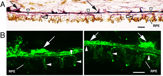
 Figure 1 of
Kochounian, Mol Vis 2016; 22:213-223.
Figure 1 of
Kochounian, Mol Vis 2016; 22:213-223.