Figure 1. One-(1P) and two-photon (2P) imaging of Hoechst 33342, autofluorescence, and Alexa-568 in the human juxtacanalicular meshwork.
1P (A–C) and 2P (D–I) fluorescence excitation imaging, at varying excitation wavelengths, resulted in unique combinations of Hoechst 33342, autofluorescence
(AF), and Alexa-568-conjugated phalloidin emission signals. Emission was captured through red (565–605 nm; A, D, G) and green (500–550 nm; B, E, H) filters. 1P excitation was at 543 nm for the red channel and 488 nm for the green channel. 2P excitation was at 750 nm (D–F) or 850 nm (G–I). Alexa-568-phalloidin fluorescence was similar in the red channel across all conditions. Hoechst 33342 nuclear fluorescence
was visible in the green channel (500–550 nm) with 2P, but not 1P, excitation, in which it was brighter at shorter excitation
wavelengths (E versus H). With 750 nm excitation (E), Hoechst 33342 was bright, but AF was almost imperceptible. With 850 nm excitation (H), AF and Hoechst 33342 intensities and visualization were more balanced. Arrows=Hoechst 33342–labeled nuclei. Asterisk=extracellular
matrix-associated AF. SP mirror=tunable prism-based spectrophotometric detector. Bar=25 μm. Insets: 2X magnification of regions
indicated by asterisks.
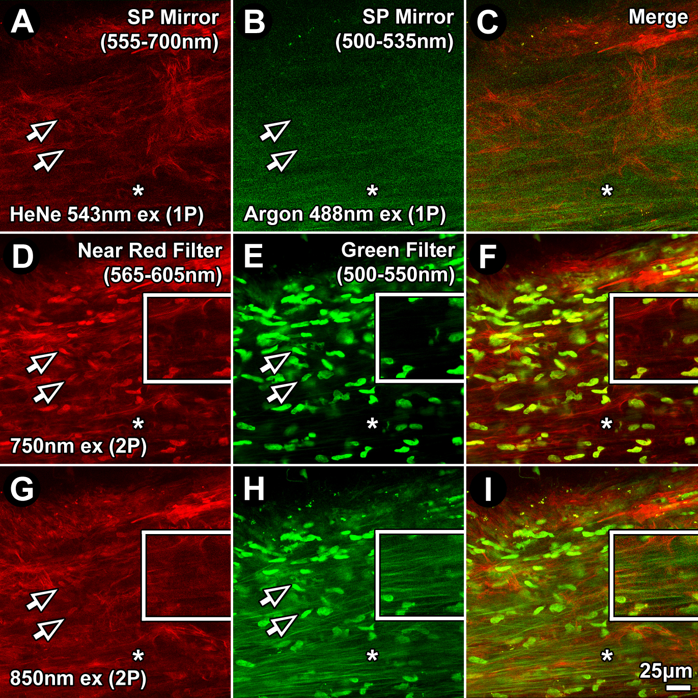
 Figure 1 of
Gonzalez, Mol Vis 2016; 22:203-212.
Figure 1 of
Gonzalez, Mol Vis 2016; 22:203-212.