Figure 5. Co-localization of the mutant EPHA2-Myc proteins with the cis-golgi apparatus in Caco-2 cells. Transiently transfected Caco-2
cells were labeled with an anti-Myc primary antibody and Cy5-conjugated secondary antibody to detect EPHA2-Myc protein (cyan),
and with an anti-GM130 primary antibody and Alexa Flour 555-conjugated secondary antibody to detect the cis-golgi apparatus
(red). Nuclei (blue) were labeled with DAPI. A: Cells expressing the EPHA2-Myc protein carrying p.P584L (second row), p.A959T (third row), or p.V972GfsX39 (fourth row)
mutation showed peripheral and cytoplasmic localization of the protein comparable to that of the wild-type EPHA2-Myc protein
(first row). The cells expressing mutant EPHA2-Myc protein carrying p.A959T and p.V972GfsX39 mutation also display some perinuclear
localization of the protein (third and fourth row). These cells show some co-localization with the cis-golgi apparatus (white
in the merged images), possibly due to the overexpression of the mutant proteins. B: Cells expressing mutant EPHA2-Myc proteins with p.T940I and p.D942fsXC71 mutations predominantly show mis-localization of
the protein in the perinuclear region and co-localization with the cis-golgi apparatus (white in the merged image in the first
and third rows; arrows). A few cells expressing these mutant proteins also exhibited peripheral and cytoplasmic localization
(second and fourth rows). Representative images from two independent experiments are shown. Scale-bar=20 µm.
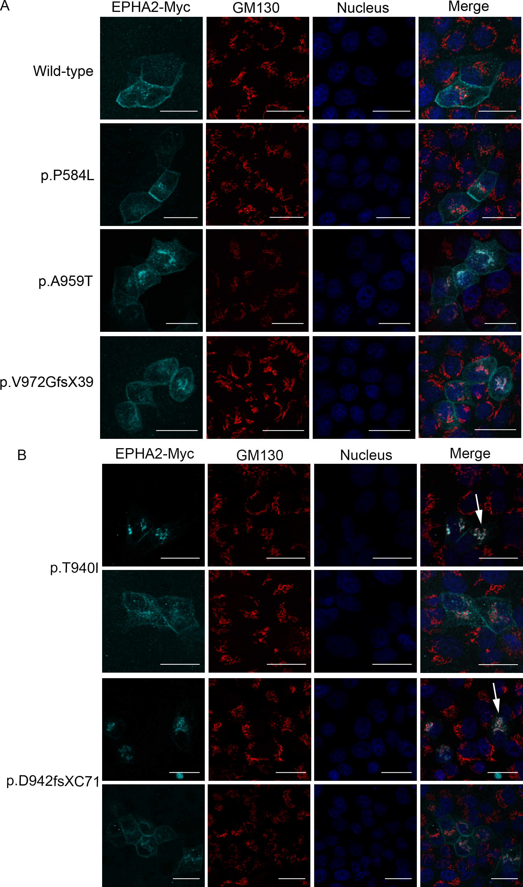
 Figure 5 of
Dave, Mol Vis 2016; 22:18-30.
Figure 5 of
Dave, Mol Vis 2016; 22:18-30.